Bone Marrow Niches and HSC Fates
A detailed reference on signaling pathways in the bone marrow and how these influence HSC fate decisions; created in partnership with Nature Reviews Immunology and Nature Reviews Molecular Cell Biology
Gao L et al. (APR 2000)
Blood 95 7 2198--203
Selective elimination of leukemic CD34(+) progenitor cells by cytotoxic T lymphocytes specific for WT1.
Hematologic malignancies such as acute and chronic myeloid leukemia are characterized by the malignant transformation of immature CD34(+) progenitor cells. Transformation is associated with elevated expression of the Wilm's tumor gene encoded transcription factor (WT1). Here we demonstrate that WT1 can serve as a target for cytotoxic T lymphocytes (CTL) with exquisite specificity for leukemic progenitor cells. HLA-A0201- restricted CTL specific for WT1 kill leukemia cell lines and inhibit colony formation by transformed CD34(+) progenitor cells isolated from patients with chronic myeloid leukemia (CML),whereas colony formation by normal CD34(+) progenitor cells is unaffected. Thus,the tissue-specific transcription factor WT1 is an ideal target for CTL-mediated purging of leukemic progenitor cells in vitro and for antigen-specific therapy of leukemia and other WT1-expressing malignancies in vivo.
View Publication
产品号#:
04535
04545
产品名:
MethoCult™ H4535 Enriched,不含EPO
MethoCult™ H4535 Enriched,不含EPO
Bruserud &O et al. (JUN 2002)
Haematologica 87 6 584--95
Leptin in human acute myelogenous leukemia: studies of in vivo levels and in vitro effects on native functional leukemia blasts.
BACKGROUND AND OBJECTIVES: Leptin receptors can be expressed by acute myelogenous leukemia (AML) cells,but the functional effects of leptin on native AML blasts have not been characterized in detail. We investigated systemic leptin levels in AML patients and in vitro effects of leptin on cultured AML blasts. DESIGN AND METHODS: Serum leptin levels were compared for patients with untreated AML and healthy controls. Native AML blasts were derived from a large group of consecutive patients,and effects of leptin on proliferation (suspension cultures and colony formation),constitutive cytokine secretion,differentiation and apoptosis regulation were assayed in vitro. RESULTS: Systemic leptin levels were decreased in patients with untreated AML,and leptin levels in acute leukemia patients were not altered during severe chemotherapy-induced cytopenia and complicating febrile neutropenia. In vitro studies demonstrated that leptin increased AML blast release of interleukin (IL) 1beta,IL6,tumor necrosis factor (TNF) alpha and granulocyte-macrophage colony-stimulating factor (GM-CSF). This enhancing effect showed no correlation with CD34 expression and was not dependent on the presence of serum,induction of differentiation or alteration of caspase 3 activity with decreased in vitro apoptosis. Leptin also increased spontaneous AML blast proliferation,whereas divergent effects on blast proliferation were observed in the presence of exogenous cytokines. The in vitro effects were usually observed at concentrations exceeding the systemic levels. INTERPRETATION AND CONCLUSIONS: Our results suggest that systemic leptin levels alone do not have a major influence on native AML blasts,but the systemic levels in combination with local leptin release in the bone marrow may affect the functional characteristics of these cells.
View Publication
产品号#:
04230
09600
09650
产品名:
MethoCult™ H4230
StemSpan™ SFEM
StemSpan™ SFEM
Alberta JA et al. (APR 2003)
Blood 101 7 2570--4
Role of the WT1 tumor suppressor in murine hematopoiesis.
The WT1 tumor-suppressor gene is expressed by many forms of acute myeloid leukemia. Inhibition of this expression can lead to the differentiation and reduced growth of leukemia cells and cell lines,suggesting that WT1 participates in regulating the proliferation of leukemic cells. However,the role of WT1 in normal hematopoiesis is not well understood. To investigate this question,we have used murine cells in which the WT1 gene has been inactivated by homologous recombination. We have found that cells lacking WT1 show deficits in hematopoietic stem cell function. Embryonic stem cells lacking WT1,although contributing efficiently to other organ systems,make only a minimal contribution to the hematopoietic system in chimeras,indicating that hematopoietic stem cells lacking WT1 compete poorly with healthy stem cells. In addition,fetal liver cells lacking WT1 have an approximately 75% reduction in erythroid blast-forming unit (BFU-E),erythroid colony-forming unit (CFU-E),and colony-forming unit-granulocyte macrophage-erythroid-megakaryocyte (CFU-GEMM). However,transplantation of fetal liver hematopoietic cells lacking WT1 will repopulate the hematopoietic system of an irradiated adult recipient in the absence of competition. We conclude that the absence of WT1 in hematopoietic cells leads to functional defects in growth potential that may be of consequence to leukemic cells that have alterations in the expression of WT1.
View Publication
产品号#:
03434
03444
产品名:
MethoCult™ GF M3434
MethoCult™ GF M3434
Gattermann N et al. (FEB 2004)
Blood 103 4 1499--502
Ineffective hematopoiesis linked with a mitochondrial tRNA mutation (G3242A) in a patient with myelodysplastic syndrome.
In a patient with refractory anemia with excess blasts (RAEB),a somatic mutation of mitochondrial transfer RNA(Leu(UUR)) was detected in bone marrow cells. Heteroduplex analysis indicated that 40% to 50% of mitochondrial DNA (mtDNA) molecules in the bone marrow (BM) carried the novel G3242A mutation. The proportion of mutant mtDNA was higher in CD34(+) cells than in the unfractionated sample. Surprisingly,the mutation was not detectable by heteroduplex analysis in the peripheral blood (PB). However,PB CD34(+) cells selected by immunomagnetic beads harbored the mutation with a proportion of approximately 50%. In hematopoietic colony assays,CD34(+) cells from BM and PB yielded only colonies with wild-type mtDNA. These results indicate that the mtDNA mutation in CD34(+) cells was associated with a maturation defect. Mitochondrial tRNA mutations impair mitochondrial protein synthesis,thereby causing dysfunction of the mitochondrial respiratory chain. We propose that this effect contributed to ineffective hematopoiesis in our patient.
View Publication
Pineault N et al. (MAR 2004)
Molecular and cellular biology 24 5 1907--17
Differential and common leukemogenic potentials of multiple NUP98-Hox fusion proteins alone or with Meis1.
NUP98-Hox fusion genes are newly identified oncogenes isolated in myeloid leukemias. Intriguingly,only Abd-B Hox genes have been reported as fusion partners,indicating that they may have unique overlapping leukemogenic properties. To address this hypothesis,we engineered novel NUP98 fusions with Hox genes not previously identified as fusion partners: the Abd-B-like gene HOXA10 and two Antennepedia-like genes,HOXB3 and HOXB4. Notably,NUP98-HOXA10 and NUP98-HOXB3 but not NUP98-HOXB4 induced leukemia in a murine transplant model,which is consistent with the reported leukemogenic potential ability of HOXA10 and HOXB3 but not HOXB4. Thus,the ability of Hox genes to induce leukemia as NUP98 fusion partners,although apparently redundant for Abd-B-like activity,is not restricted to this group,but rather is determined by the intrinsic leukemogenic potential of the Hox partner. We also show that the potent leukemogenic activity of Abd-B-like Hox genes is correlated with their strong ability to block hematopoietic differentiation. Conversely,coexpression of the Hox cofactor Meis1 alleviated the requirement of a strong intrinsic Hox-transforming potential to induce leukemia. Our results support a model in which many if not all Hox genes can be leukemogenic and point to striking functional overlap not previously appreciated,presumably reflecting common regulated pathways.
View Publication

 EasySep™小鼠TIL(CD45)正选试剂盒


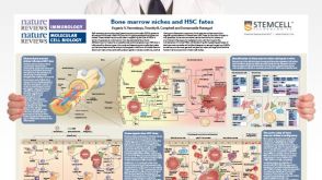 挂图Bone Marrow Niches and HSC Fates A detailed reference on signaling pathways in the bone marrow and how these influence HSC fate decisions; created in partnership with Nature Reviews Immunology and Nature Reviews Molecular Cell Biology


 沪公网安备31010102008431号