Kolly L et al. (SEP 2009)
Journal of immunology (Baltimore,Md. : 1950) 183 6 4003--12
Inflammatory role of ASC in antigen-induced arthritis is independent of caspase-1, NALP-3, and IPAF.
Because IL-1beta plays an important role in inflammation in human and murine arthritis,we investigated the contribution of the inflammasome components ASC,NALP-3,IPAF,and caspase-1 to inflammatory arthritis. We first studied the phenotype of ASC-deficient and wild-type mice during Ag-induced arthritis (AIA). ASC(-/-) mice showed reduced severity of AIA,decreased levels of synovial IL-1beta,and diminished serum amyloid A levels. In contrast,mice deficient in NALP-3,IPAF,or caspase-1 did not show any alteration of joint inflammation,thus indicating that ASC associated effects on AIA are independent of the classical NALP-3 or IPAF inflammasomes. Because ASC is a ubiquitous cytoplasmic protein that has been implicated in multiple cellular processes,we explored other pathways through which ASC may modulate inflammation. Ag-specific proliferation of lymph node and spleen cells from ASC-deficient mice was significantly decreased in vitro,as was the production of IFN-gamma,whereas IL-10 production was enhanced. TCR ligation by anti-CD3 Abs in the presence or absence of anti-CD28 Abs induced a reduction in T cell proliferation in ASC(-/-) T cells compared with wild-type ones. In vivo lymph node cell proliferation was also significantly decreased in ASC(-/-) mice,but no effects on apoptosis were observed either in vitro or in vivo in these mice. In conclusion,these results strongly suggest that ASC modulates joint inflammation in AIA through its effects on cell-mediated immune responses but not via its implication in inflammasome formation.
View Publication
文献
Marks BR et al. (OCT 2009)
Nature immunology 10 10 1125--32
Thymic self-reactivity selects natural interleukin 17-producing T cells that can regulate peripheral inflammation.
Interleukin 17 (IL-17)-producing CD4(+) helper T cells (T(H)-17 cells) share a developmental relationship with Foxp3(+) regulatory T cells (T(reg) cells). Here we show that a T(H)-17 population differentiates in the thymus in a manner influenced by recognition of self antigen and by the cytokines IL-6 and transforming growth factor-beta (TGF-beta). Like previously described T(H)-17 cells,the T(H)-17 cells that developed in the thymus expressed the transcription factor RORgamma t and the IL-23 receptor. These cells also expressed alpha(4)beta(1) integrins and the chemokine receptor CCR6 and were recruited to the lung,gut and liver. In the liver,these cells secreted IL-22 in response to self antigen and mediated host protection during inflammation. Thus,T(H)-17 cells,like T(reg) cells,can be selected by self antigens in the thymus.
View Publication
文献
Su X et al. (FEB 2010)
Journal of immunology (Baltimore,Md. : 1950) 184 3 1630--41
Tumor microenvironments direct the recruitment and expansion of human Th17 cells.
Although Th17 cells play critical roles in the pathogenesis of many inflammatory and autoimmune diseases,their prevalence among tumor-infiltrating lymphocytes (TILs) and function in human tumor immunity remains largely unknown. We have recently demonstrated high percentages of Th17 cells in TILs from ovarian cancer patients,but the mechanisms of accumulation of these Th17 cells in the tumor microenvironment are still unclear. In this study,we further showed elevated Th17 cell populations in the TILs obtained from melanoma and breast and colon cancers,suggesting that development of tumor-infiltrating CD4(+) Th17 cells may be a general feature in cancer patients. We then demonstrated that tumor microenvironmental RANTES and MCP-1 secreted by tumor cells and tumor-derived fibroblasts mediate the recruitment of Th17 cells. In addition to their recruitment,we found that tumor cells and tumor-derived fibroblasts produce a proinflammatory cytokine milieu as well as provide cell-cell contact engagement that facilitates the generation and expansion of Th17 cells. We also showed that inflammatory TLR and nucleotide oligomerization binding domain 2 signaling promote the attraction and generation of Th17 cells induced by tumor cells and tumor-derived fibroblasts. These results identify Th17 cells as an important component of human TILs,demonstrate mechanisms involved in the recruitment and regulation of Th17 cells in tumor microenvironments,and provide new insights relevant for the development of novel cancer immunotherapeutic approaches.
View Publication
文献
Fang Y et al. (JUN 2010)
Journal of leukocyte biology 87 6 1019--28
Comparison of sensitivity of Th1, Th2, and Th17 cells to Fas-mediated apoptosis.
Following activation through the TCR,CD4+ T cells can differentiate into three major subsets: Th1,Th2,and Th17 cells. IL-17-secreting Th17 cells play an important role in the pathogenesis of several autoimmune diseases and in immune responses to pathogens,but little is known about the regulation of apoptosis in Th17 cells. In this study,the sensitivity of in vitro-polarized Th1,Th2,and Th17 cells to Fas-mediated apoptosis was compared directly by different methods. The order of sensitivity of T cell subsets to Fas-mediated apoptosis is: Th1 textgreater Th17 textgreater Th2. The greater sensitivity of Th17 cells to Fas-mediated apoptosis compared with Th2 cells correlated with their higher expression of FasL and comparable expression of the antiapoptotic molecule FLIP. The decreased sensitivity of Th17 compared with Th1 cells correlated with the higher expression of FLIP by Th17 cells. Transgenic overexpression of FLIP in T cells protected all three subsets from Fas-mediated apoptosis. These findings provide new knowledge for understanding how survival of different subsets of T cells is regulated.
View Publication
文献
Hale JS et al. (JUN 2010)
Journal of immunology (Baltimore,Md. : 1950) 184 11 5964--8
Cutting Edge: Rag deletion in peripheral T cells blocks TCR revision.
Mature CD4(+)Vbeta5(+) T cells that recognize a peripherally expressed endogenous superantigen are tolerized either by deletion or TCR revision. In Vbeta5 transgenic mice,this latter tolerance pathway results in the appearance of CD4(+)Vbeta5(-)TCRbeta(+) T cells,coinciding with Rag1,Rag2,and TdT expression and the accumulation of V(beta)-DJ(beta) recombination intermediates in peripheral CD4(+) T cells. Because postthymic RAG-dependent TCR rearrangement has remained controversial,we sought to definitively determine whether TCR revision is an extrathymic process that occurs in mature peripheral T cells. We show in this study that Rag deletion in post-positive selection T cells in Vbeta5 transgenic mice blocks TCR revision in vivo and that mature peripheral T cells sorted to remove cells bearing endogenous TCRbeta-chains can express newly generated TCRbeta molecules in adoptive hosts. These findings unambiguously demonstrate postthymic,RAG-dependent TCR rearrangement and define TCR revision as a tolerance pathway that targets mature peripheral CD4(+) T cells.
View Publication
文献
Poholek AC et al. (JUL 2010)
Journal of immunology (Baltimore,Md. : 1950) 185 1 313--26
In vivo regulation of Bcl6 and T follicular helper cell development.
Follicular helper T (T(FH)) cells,defined by expression of the surface markers CXCR5 and programmed death receptor-1 (PD-1) and synthesis of IL-21,require upregulation of the transcriptional repressor Bcl6 for their development and function in B cell maturation in germinal centers. We have explored the role of B cells and the cytokines IL-6 and IL-21 in the in vivo regulation of Bcl6 expression and T(FH) cell development. We found that T(FH) cells are characterized by a Bcl6-dependent downregulation of P-selectin glycoprotein ligand 1 (PSGL1,a CCL19- and CCL21-binding protein),indicating that,like CXCR5 and PD-1 upregulation,modulation of PSGL1 expression is part of the T(FH) cell program of differentiation. B cells were neither required for initial upregulation of Bcl6 nor PSGL1 downregulation,suggesting these events preceded T-B cell interactions,although they were required for full development of the T(FH) cell phenotype,including CXCR5 and PD-1 upregulation,and IL-21 synthesis. Bcl6 upregulation and T(FH) cell differentiation were independent of IL-6 and IL-21,revealing that either cytokine is not absolutely required for development of Bcl6(+) T(FH) cells in vivo. These data increase our understanding of Bcl6 regulation in T(FH) cells and their differentiation in vivo and identifies a new surface marker that may be functionally relevant in this subset.
View Publication
文献
Carr EL et al. (JUL 2010)
Journal of immunology (Baltimore,Md. : 1950) 185 2 1037--44
Glutamine uptake and metabolism are coordinately regulated by ERK/MAPK during T lymphocyte activation.
Activation of a naive T cell is a highly energetic event,which requires a substantial increase in nutrient metabolism. Upon stimulation,T cells increase in size,rapidly proliferate,and differentiate,all of which lead to a high demand for energetic and biosynthetic precursors. Although amino acids are the basic building blocks of protein biosynthesis and contribute to many other metabolic processes,the role of amino acid metabolism in T cell activation has not been well characterized. We have found that glutamine in particular is required for T cell function. Depletion of glutamine blocks proliferation and cytokine production,and this cannot be rescued by supplying biosynthetic precursors of glutamine. Correlating with the absolute requirement for glutamine,T cell activation induces a large increase in glutamine import,but not glutamate import,and this increase is CD28-dependent. Activation coordinately enhances expression of glutamine transporters and activities of enzymes required to allow the use of glutamine as a Krebs cycle substrate in T cells. The induction of glutamine uptake and metabolism requires ERK function,providing a link to TCR signaling. Together,these data indicate that regulation of glutamine use is an important component of T cell activation. Thus,a better understanding of glutamine sensing and use in T cells may reveal novel targets for immunomodulation.
View Publication
文献
Da Silva CA et al. (DEC 2010)
American journal of respiratory and critical care medicine 182 12 1482--91
Chitin particles are multifaceted immune adjuvants.
RATIONALE: Chitin is a ubiquitous polysaccharide in fungi,insects,allergens,and parasites that is released at sites of infection. Its role in the generation of tissue inflammation,however,is not fully understood. OBJECTIVES: We hypothesized that chitin is an important adjuvant for adaptive immunity. METHODS: Mice were injected with a solution of ovalbumin and chitin. MEASUREMENTS AND MAIN RESULTS: We used in vivo and ex vivo/in vitro approaches to characterize the ability of chitin fragments to foster adaptive immune responses against ovalbumin and compared these responses to those induced by aluminum hydroxide (alum). In vivo,ovalbumin challenge caused an eosinophil-rich pulmonary inflammatory response,Th2 cytokine elaboration,IgE induction,and mucus metaplasia in mice that had been sensitized with ovalbumin plus chitin or ovalbumin plus alum. Toll-like receptor-2,MyD88,and IL-17A played critical roles in the chitin-induced responses,and MyD88 and IL-17A played critical roles in the alum-induced responses. In vitro,CD4(+) T cells from mice sensitized with ovalbumin plus chitin were incubated with ovalbumin-stimulated bone marrow-derived dendritic cells. In these experiments,CD4(+) T-cell proliferation,IL-5,IL-13,IFN-γ,and IL-17A production were appreciated. Toll-like receptor-2,MyD88,and IL-17A played critical roles in these in vitro adjuvant properties of chitin. TLR-2 was required for cell proliferation,whereas IL-17 and TLR-2 were required for cytokine elaboration. IL-17A also inhibited the generation of adaptive Th1 responses. CONCLUSIONS: These studies demonstrate that chitin is a potent multifaceted adjuvant that induces adaptive Th2,Th1,and Th17 immune responses. They also demonstrate that the adjuvant properties of chitin are mediated by a pathway(s) that involves and is regulated by TLR-2,MyD88,and IL-17A.
View Publication
文献
Quintarelli C et al. (MAR 2011)
Blood 117 12 3353--62
High-avidity cytotoxic T lymphocytes specific for a new PRAME-derived peptide can target leukemic and leukemic-precursor cells.
The cancer testis antigen (CTA) preferentially expressed antigen of melanoma (PRAME) is overexpressed by many hematologic malignancies,but is absent on normal tissues,including hematopoietic progenitor cells,and may therefore be an appropriate candidate for T cell-mediated immunotherapy. Because it is likely that an effective antitumor response will require high-avidity,PRAME-specific cytotoxic T lymphocytes (CTLs),we attempted to generate such CTLs using professional and artificial antigen-presenting cells loaded with a peptide library spanning the entire PRAME protein and consisting of 125 synthetic pentadecapeptides overlapping by 11 amino acids. We successfully generated polyclonal,PRAME-specific CTL lines and elicited high-avidity CTLs,with a high proportion of cells recognizing a previously uninvestigated HLA-A*02-restricted epitope,P435-9mer (NLTHVLYPV). These PRAME-CTLs could be generated both from normal donors and from subjects with PRAME(+) hematologic malignancies. The cytotoxic activity of our PRAME-specific CTLs was directed not only against leukemic blasts,but also against leukemic progenitor cells as assessed by colony-forming-inhibition assays,which have been implicated in leukemia relapse. These PRAME-directed CTLs did not affect normal hematopoietic progenitors,indicating that this approach may be of value for immunotherapy of PRAME(+) hematologic malignancies.
View Publication
挂图
Human Immune Cytokines
Infographic of key cytokines for expansion, differentiation and characterization of major immune cell types
文献
Aghaeepour N et al. (AUG 2017)
Journal of immunology (Baltimore,Md. : 1950)
Deep Immune Profiling of an Arginine-Enriched Nutritional Intervention in Patients Undergoing Surgery.
Application of high-content immune profiling technologies has enormous potential to advance medicine. Whether these technologies reveal pertinent biology when implemented in interventional clinical trials is an important question. The beneficial effects of preoperative arginine-enriched dietary supplements (AES) are highly context specific,as they reduce infection rates in elective surgery,but possibly increase morbidity in critically ill patients. This study combined single-cell mass cytometry with the multiplex analysis of relevant plasma cytokines to comprehensively profile the immune-modifying effects of this much-debated intervention in patients undergoing surgery. An elastic net algorithm applied to the high-dimensional mass cytometry dataset identified a cross-validated model consisting of 20 interrelated immune features that separated patients assigned to AES from controls. The model revealed wide-ranging effects of AES on innate and adaptive immune compartments. Notably,AES increased STAT1 and STAT3 signaling responses in lymphoid cell subsets after surgery,consistent with enhanced adaptive mechanisms that may protect against postsurgical infection. Unexpectedly,AES also increased ERK and P38 MAPK signaling responses in monocytic myeloid-derived suppressor cells,which was paired with their pronounced expansion. These results provide novel mechanistic arguments as to why AES may exert context-specific beneficial or adverse effects in patients with critical illness. This study lays out an analytical framework to distill high-dimensional datasets gathered in an interventional clinical trial into a fairly simple model that converges with known biology and provides insight into novel and clinically relevant cellular mechanisms.
View Publication
文献
Zhang Y et al. ( 2018)
Nature communications 9 1 6
Nanoparticle anchoring targets immune agonists to tumors enabling anti-cancer immunity without systemic toxicity.
Immunostimulatory agents such as agonistic anti-CD137 and interleukin (IL)-2 generate effective anti-tumor immunity but also elicit serious toxicities,hampering their clinical application. Here we show that combination therapy with anti-CD137 and an IL-2-Fc fusion achieves significant initial anti-tumor activity,but also lethal immunotoxicity deriving from stimulation of circulating leukocytes. To overcome this toxicity,we demonstrate that anchoring IL-2 and anti-CD137 on the surface of liposomes allows these immune agonists to rapidly accumulate in tumors while lowering systemic exposure. In multiple tumor models,immunoliposome delivery achieves anti-tumor activity equivalent to free IL-2/anti-CD137 but with the complete absence of systemic toxicity. Immunoliposomes stimulated tumor infiltration by cytotoxic lymphocytes,cytokine production,and granzyme expression,demonstrating equivalent immunostimulatory effects to the free drugs in the local tumor microenvironment. Thus,surface-anchored particle delivery may provide a general approach to exploit the potent stimulatory activity of immune agonists without debilitating systemic toxicities.
View Publication

 EasySep™小鼠TIL(CD45)正选试剂盒


 文献
文献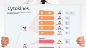 挂图Human Immune Cytokines Infographic of key cytokines for expansion, differentiation and characterization of major immune cell types
 沪公网安备31010102008431号