HIV disease progression despite suppression of viral replication is associated with exhaustion of lymphopoiesis.
The mechanisms of CD4(+) T-cell count decline,the hallmark of HIV disease progression,and its relationship to elevated levels of immune activation are not fully understood. Massive depletion of CD4(+) T cells occurs during the course of HIV-1 infection,so that maintenance of adequate CD4(+) T-cell levels probably depends primarily on the capacity to renew depleted lymphocytes,that is,the lymphopoiesis. We performed here a comprehensive study of quantitative and qualitative attributes of CD34(+) hematopoietic progenitor cells directly from the blood of a large set of HIV-infected persons compared with uninfected donors,in particular the elderly. Our analyses underline a marked impairment of primary immune resources with the failure to maintain adequate lymphocyte counts. Systemic immune activation emerges as a major correlate of altered lymphopoiesis,which can be partially reversed with prolonged antiretroviral therapy. Importantly,HIV disease progression despite elite control of HIV replication or virologic success on antiretroviral treatment is associated with persistent damage to the lymphopoietic system or exhaustion of lymphopoiesis. These findings highlight the importance of primary hematopoietic resources in HIV pathogenesis and the response to antiretroviral treatments.
View Publication
文献
Shi X et al. (MAY 2011)
Infection and immunity 79 5 2031--42
Thymopoietic and bone marrow response to murine Pneumocystis pneumonia.
CD4(+) T cells play a key role in host defense against Pneumocystis infection. To define the role of naïve CD4(+) T cell production through the thymopoietic response in host defense against Pneumocystis infection,Pneumocystis murina infection in the lung was induced in adult male C57BL/6 mice with and without prior thymectomy. Pneumocystis infection caused a significant increase in the number of CCR9(+) multipotent progenitor (MPP) cells in the bone marrow and peripheral circulation,an increase in populations of earliest thymic progenitors (ETPs) and double negative (DN) thymocytes in the thymus,and recruitment of naïve and total CD4(+) T cells into the alveolar space. The level of murine signal joint T cell receptor excision circles (msjTRECs) in spleen CD4(+) cells was increased at 5 weeks post-Pneumocystis infection. In thymectomized mice,the numbers of naïve,central memory,and total CD4(+) T cells in all tissues examined were markedly reduced following Pneumocystis infection. This deficiency of naïve and central memory CD4(+) T cells was associated with delayed pulmonary clearance of Pneumocystis. Extracts of Pneumocystis resulted in an increase in the number of CCR9(+) MPPs in the cultured bone marrow cells. Stimulation of cultured bone marrow cells with ligands to Toll-like receptor 2 ([TLR-2] zymosan) and TLR-9 (ODN M362) each caused a similar increase in CCR9(+) MPP cells via activation of the Jun N-terminal protein kinase (JNK) pathway. These results demonstrate that enhanced production of naïve CD4(+) T lymphocytes through the thymopoietic response and enhanced delivery of lymphopoietic precursors from the bone marrow play an important role in host defense against Pneumocystis infection.
View Publication
文献
Vieillard V et al. (AUG 2005)
Proceedings of the National Academy of Sciences 102 31 10981--86
NK cytotoxicity against CD4+ T cells during HIV-1 infection: A gp41 peptide induces the expression of an NKp44 ligand
HIV infection leads to a state of chronic immune activation and progressive deterioration in immune function,manifested most recognizably by the progressive depletion of CD4+ T cells. A substantial percentage of natural killer (NK) cells from patients with HIV infection are activated and express the natural cytotoxicity receptor (NCR) NKp44. Here we show that a cellular ligand for NKp44 (NKp44L) is expressed during HIV-1 infection and is correlated with both the progression of CD4+ T cell depletion and the increase of viral load. CD4+ T cells expressing this ligand are highly sensitive to the NK lysis activity mediated by NKp44+ NK cells. The expression of NKp44L is induced by the linear motif NH2-SWSNKS-COOH of the HIV-1 envelope gp41 protein. This highly conserved motif appears critical to the sharp increase in NK lysis of CD4+ T cells from HIV-infected patients. These studies strongly suggest that induction of NKp44L plays a key role in the lysis of CD4+ T cells by activated NK cells in HIV infection and consequently provide a framework for considering how HIV-1 may use NK cell immune surveillance to trigger CD4+ T cells. Understanding this mechanism may help to develop future therapeutic strategies and vaccines against HIV-1 infection.
View Publication
文献
Abdelwahab SF et al. (DEC 2003)
Proceedings of the National Academy of Sciences of the United States of America 100 25 15006--10
HIV-1-suppressive factors are secreted by CD4+ T cells during primary immune responses.
CD4+ T cells are required for immunity against many viral infections,including HIV-1 where a positive correlation has been observed between strong recall responses and low HIV-1 viral loads. Some HIV-1-specific CD4+ T cells are preferentially infected with HIV-1,whereas others escape infection by unknown mechanisms. One possibility is that some CD4+ T cells are protected from infection by the secretion of soluble HIV-suppressive factors,although it is not known whether these factors are produced during primary antigen-specific responses. Here,we show that soluble suppressive factors are produced against CXCR4 and CCR5 isolates of HIV-1 during the primary immune response of human CD4+ T cells. This activity requires antigenic stimulation of naïve CD4+ T cells. One anti-CXCR4 factor is macrophage-derived chemokine (chemokine ligand 22,CCL22),and anti-CCR5 factors include macrophage inflammatory protein-1 alpha (CCL3),macrophage inflammatory protein-1 beta (CCL4),and RANTES (regulated upon activation of normal T cells expressed and secreted) (CCL5). Intracellular staining confirms that CD3+CD4+ T cells are the source of the prototype HIV-1-inhibiting chemokines CCL22 and CCL4. These results show that CD4+ T cells secrete an evolving HIV-1-suppressive activity during the primary immune response and that this activity is comprised primarily of CC chemokines. The data also suggest that production of such factors should be considered in the design of vaccines against HIV-1 and as a mechanism whereby the host can control infections with this virus.
View Publication
文献
Jones DC et al. (JUL 2003)
Journal of immunology 171 1 196--203
Peroxisome proliferator-activated receptor alpha negatively regulates T-bet transcription through suppression of p38 mitogen-activated protein kinase activation.
Expression of the nuclear hormone receptor peroxisome proliferator-activated receptor alpha (PPARalpha) in resting lymphocytes was recently established,although the physiologic role(s) played by this nuclear hormone receptor in these cell types remains unresolved. In this study,we used CD4(+) T cells isolated from PPARalpha(-/-) and wild-type mice,as well as cell lines that constitutively express PPARalpha,in experiments designed to evaluate the role of this hormone receptor in the regulation of T cell function. We report that activated CD4(+) T cells lacking PPARalpha produce increased levels of IFN-gamma,but significantly lower levels of IL-2 when compared with activated wild-type CD4(+) T cells. Furthermore,we demonstrate that PPARalpha regulates the expression of these cytokines by CD4(+) T cells in part,through its ability to negatively regulate the transcription of T-bet. The induction of T-bet expression in CD4(+) T cells was determined to be positively influenced by p38 mitogen-activated protein (MAP) kinase activation,and the presence of unliganded PPARalpha effectively suppressed the phosphorylation of p38 MAP kinase. The activation of PPARalpha with highly specific ligands relaxed its capacity to suppress p38 MAP kinase phosphorylation and promoted T-bet expression. These results demonstrate a novel DNA-binding independent and agonist-controlled regulatory influence by the nuclear hormone receptor PPARalpha.
View Publication
文献
Schlecht G et al. (OCT 2001)
Journal of immunology (Baltimore,Md. : 1950) 167 8 4215--21
Induction of CTL and nonpolarized Th cell responses by CD8alpha(+) and CD8alpha(-) dendritic cells.
Two distinct dendritic cell (DC) subpopulations have been evidenced in mice on the basis of their differential CD8alpha expression and their localization in lymphoid organs. Several reports suggest that CD8alpha(+) and CD8alpha(-) DC subsets could be functionally different. In this study,using a panel of MHC class I- and/or class II-restricted peptides,we analyzed CD4(+) and CD8(+) T cell responses obtained after i.v. injection of freshly purified peptide-pulsed DC subsets. First,we showed that both DC subsets efficiently induce specific CTL responses and Th1 cytokine production in the absence of CD4(+) T cell priming. Second,we showed that in vivo activation of CD4(+) T cells by CD8alpha(+) or CD8alpha(-) DC,injected i.v.,leads to a nonpolarized Th response with production of both Th1 and Th2 cytokines. The CD8alpha(-) subset induced a higher production of Th2 cytokines such as IL-4 and IL-10 than the CD8alpha(+) subset. However,IL-5 was produced by CD4(+) T cells activated by both DC subsets. When both CD4(+) and CD8(+) T cells were primed by DC injected i.v.,a similar pattern of cytokines was observed,but,under these conditions,Th1 cytokines were mainly produced by CD8(+) T cells,while Th2 cytokines were produced by CD4(+) T cells. Thus,this study clearly shows that CD4(+) T cell responses do not influence the development of specific CD8(+) T cell cytotoxic responses induced either by CD8alpha(+) or CD8alpha(-) DC subsets.
View Publication

 EasySep™小鼠TIL(CD45)正选试剂盒


 文献
文献
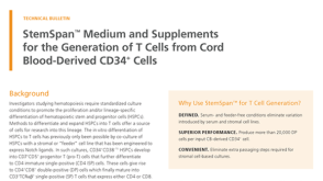 技术公告StemSpan™ Medium and Supplements for the Generation of T Cells from Cord Blood-Derived CD34+ Cells
 Immunology Features
 1:23
How to Isolate Cells with EasySep™ Column-Free Cell Separation Technology
1:23
How to Isolate Cells with EasySep™ Column-Free Cell Separation Technology 产品手册Tools for Your Immunology Research