Binder LI et al. (SEP 1984)
Proceedings of the National Academy of Sciences of the United States of America 81 17 5613--7
Heterogeneity of microtubule-associated protein 2 during rat brain development.
The electrophoretic pattern of the large microtubule-associated protein,MAP2,changes during rat brain development. Immunoblots of NaDodSO4 extracts obtained from the cerebral cortex,cerebellum,and thalamus at 10-15 days after birth reveal only a single electrophoretic species when probed with any of three MAP2 monoclonal antibodies. By contrast,adult MAP2 contains two immunoreactive species,MAP2a and MAP2b. The single band of MAP2 from immature brain electrophoretically comigrates with adult MAP2b. Between postnatal days 17 and 18,immature MAP2 simultaneously resolves into two species in both the cerebellum and cerebral cortex. Immunoblots of NaDodSO4 extracts from spinal cord demonstrate the adult complement of MAP2 by day 10,indicating that MAP2 does not change coordinately throughout the entire central nervous system. In vitro cAMP-dependent phosphorylation of immature MAP2 causes a band split reminiscent of that seen during brain development in vivo. The possibility that the developmentally regulated changes observed in MAP2 during brain maturation are due to timed phosphorylation events is discussed.
View Publication
Organic cation transporter-mediated ergothioneine uptake in mouse neural progenitor cells suppresses proliferation and promotes differentiation into neurons.
The aim of the present study is to clarify the functional expression and physiological role in neural progenitor cells (NPCs) of carnitine/organic cation transporter OCTN1/SLC22A4,which accepts the naturally occurring food-derived antioxidant ergothioneine (ERGO) as a substrate in vivo. Real-time PCR analysis revealed that mRNA expression of OCTN1 was much higher than that of other organic cation transporters in mouse cultured cortical NPCs. Immunocytochemical analysis showed colocalization of OCTN1 with the NPC marker nestin in cultured NPCs and mouse embryonic carcinoma P19 cells differentiated into neural progenitor-like cells (P19-NPCs). These cells exhibited time-dependent [(3)H]ERGO uptake. These results demonstrate that OCTN1 is functionally expressed in murine NPCs. Cultured NPCs and P19-NPCs formed neurospheres from clusters of proliferating cells in a culture time-dependent manner. Exposure of cultured NPCs to ERGO or other antioxidants (edaravone and ascorbic acid) led to a significant decrease in the area of neurospheres with concomitant elimination of intracellular reactive oxygen species. Transfection of P19-NPCs with small interfering RNA for OCTN1 markedly promoted formation of neurospheres with a concomitant decrease of [(3)H]ERGO uptake. On the other hand,exposure of cultured NPCs to ERGO markedly increased the number of cells immunoreactive for the neuronal marker βIII-tubulin,but decreased the number immunoreactive for the astroglial marker glial fibrillary acidic protein (GFAP),with concomitant up-regulation of neuronal differentiation activator gene Math1. Interestingly,edaravone and ascorbic acid did not affect such differentiation of NPCs,in contrast to the case of proliferation. Knockdown of OCTN1 increased the number of cells immunoreactive for GFAP,but decreased the number immunoreactive for βIII-tubulin,with concomitant down-regulation of Math1 in P19-NPCs. Thus,OCTN1-mediated uptake of ERGO in NPCs inhibits cellular proliferation via regulation of oxidative stress,and also promotes cellular differentiation by modulating the expression of basic helix-loop-helix transcription factors via an unidentified mechanism different from antioxidant action.
View Publication
产品号#:
05707
产品名:
NeuroCult™化学解离试剂盒(小鼠)
Jackson TC et al. (FEB 2018)
Experimental Neurology 300 232--246
BrainPhys increases neurofilament levels in CNS cultures, and facilitates investigation of axonal damage after a mechanical stretch-injury in vitro
Neurobasal®/B27 is a gold standard culture media used to study primary neurons in vitro. An alternative media (BrainPhys®/SM1) was recently developed which robustly enhances neuronal activity vs. Neurobasal® or DMEM. To the best of our knowledge BrainPhys® has not been explored in the setting of neuronal injury. Here we characterized the utility of BrainPhys® in a model of in vitro mechanical-stretch injury. METHODS/RESULTSPrimary rat cortical neurons were maintained in classic Neurobasal®,or sequentially maintained in Neurocult® followed by BrainPhys® (hereafter simply referred to as BrainPhys® maintained neurons?). The levels of axonal markers and proteins involved in neurotransmission were compared on day in vitro 10 (DIV10). BrainPhys® maintained neurons had higher levels of GluN2B,GluR1,Neurofilament light/heavy chain (NF-L & NF-H),and protein phosphatase 2 A (PP2A) vs. neurons in Neurobasal®. Mechanical stretch-injury (50ms/54% biaxial stretch) to BrainPhys® maintained neurons modestly (albeit significantly) increased 24h lactate dehydrogenase (LDH) levels but markedly decreased axonal NF-L levels post-injury vs. uninjured controls or neurons given a milder 38% stretch-injury. Furthermore,two 54% stretch-injuries (in tandem) exacerbated 24h LDH release,increased α-spectrin breakdown products (SBDPs),and decreased Tau levels. Also,BrainPhys® maintained cultures had decreased markers of cell damage 24h after a single 54% stretch-injury vs. neurons in Neurobasal®. Finally,we tested the hypothesis that lentivirus mediated overexpression of the pro-death protein RBM5 exacerbates neuronal and/or axonal injury in primary CNS cultures. RBM5 overexpression vs. empty-vector controls increased 24h LDH release,and SBDP levels,after a single 54% stretch-injury but did not affect NF-L levels or Tau. CONCLUSIONBrainPhys® is a promising new reagent which facilities the investigation of molecular targets involved in axonal and/or neuronal injury in vitro.
View Publication
产品号#:
05790
05792
05793
05794
05795
产品名:
BrainPhys™神经元培养基
BrainPhys™神经元培养基和SM1试剂盒
BrainPhys™ 神经元培养基N2-A和SM1试剂盒
BrainPhys™原代神经元试剂盒
BrainPhys™ hPSC 神经元试剂盒
Jessick VJ et al. ( 2013)
International journal of physiology,pathophysiology and pharmacology 5 4 216--27
Investigating the role of the actin regulating complex ARP2/3 in rapid ischemic tolerance induced neuro-protection.
Neuronal morphology is highly sensitive to ischemia,although some re-organization may promote neuroprotection. In this study we investigate the role of actin regulating proteins (ARP2,ARP3 and WAVE-1) and their role in rapid ischemic tolerance. Using an established in vitro model of rapid ischemic tolerance,we show that WAVE-1 protein levels are stabilized following brief tolerance inducing ischemia (preconditioning). The stabilization appears to be due to a reduction in the ubiquitination of WAVE-1. Levels of ARP2,ARP3 and N-WASP were not affected by ischemic preconditioning. Immunocytochemical studies show a relocalization of ARP2 and ARP3 proteins in neurons following preconditioning ischemia,as well as a re-organization of actin. Blocking the protein kinase CK2 using emodin blocks ischemic tolerance,and our data suggests CK2 binds to WAVE-1 in neurons. We observe an increase in binding of the ARP2 subunit with WAVE-1. The neuroprotection observed following preconditioning is inhibited when cells are transduced with an N-WASP CA domain that blocks the activation of ARP2/3. Together these data show that ischemia affects actin regulating enzymes,and that the ARP2/3 pathway plays a role in rapid ischemic tolerance induced neuroprotection.
View Publication


 EasySep™小鼠TIL(CD45)正选试剂盒




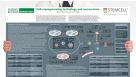 挂图Cell-Reprogramming Technology and Neuroscience Details on human iPSC-derived models of neuropsychiatric and neurodegenerative disorders


 沪公网安备31010102008431号