Lee S-HH et al. (JUN 2000)
Nature biotechnology 18 6 675--9
Efficient generation of midbrain and hindbrain neurons from mouse embryonic stem cells.
Embryonic stem (ES) cells are clonal cell lines derived from the inner cell mass of the developing blastocyst that can proliferate extensively in vitro and are capable of adopting all the cell fates in a developing embryo. Clinical interest in the use of ES cells has been stimulated by studies showing that isolated human cells with ES properties from the inner cell mass or developing germ cells can provide a source of somatic precursors. Previous studies have defined in vitro conditions for promoting the development of specific somatic fates,specifically,hematopoietic,mesodermal,and neurectodermal. In this study,we present a method for obtaining dopaminergic (DA) and serotonergic neurons in high yield from mouse ES cells in vitro. Furthermore,we demonstrate that the ES cells can be obtained in unlimited numbers and that these neuron types are generated efficiently. We generated CNS progenitor populations from ES cells,expanded these cells and promoted their differentiation into dopaminergic and serotonergic neurons in the presence of mitogen and specific signaling molecules. The differentiation and maturation of neuronal cells was completed after mitogen withdrawal from the growth medium. This experimental system provides a powerful tool for analyzing the molecular mechanisms controlling the functions of these neurons in vitro and in vivo,and potentially for understanding and treating neurodegenerative and psychiatric diseases.
View Publication
产品号#:
06902
06952
07152
07157
00321
00322
00323
00324
00325
产品名:
N2 添加物-A
Stanurova J et al. (AUG 2016)
Scientific reports 6 August 30792
Angelman syndrome-derived neurons display late onset of paternal UBE3A silencing.
Genomic imprinting is an epigenetic phenomenon resulting in parent-of-origin-specific gene expression that is regulated by a differentially methylated region. Gene mutations or failures in the imprinting process lead to the development of imprinting disorders,such as Angelman syndrome. The symptoms of Angelman syndrome are caused by the absence of functional UBE3A protein in neurons of the brain. To create a human neuronal model for Angelman syndrome,we reprogrammed dermal fibroblasts of a patient carrying a defined three-base pair deletion in UBE3A into induced pluripotent stem cells (iPSCs). In these iPSCs,both parental alleles are present,distinguishable by the mutation,and express UBE3A. Detailed characterization of these iPSCs demonstrated their pluripotency and exceptional stability of the differentially methylated region regulating imprinted UBE3A expression. We observed strong induction of SNHG14 and silencing of paternal UBE3A expression only late during neuronal differentiation,in vitro. This new Angelman syndrome iPSC line allows to study imprinted gene regulation on both parental alleles and to dissect molecular pathways affected by the absence of UBE3A protein.
View Publication
产品号#:
05850
05857
05870
05875
07174
85850
85857
85870
85875
100-0485
100-1077
产品名:
mTeSR™1
mTeSR™1
温和细胞解离试剂
ReLeSR™
Brohawn DG et al. (AUG 2016)
PloS one 11 8 e0160520
RNAseq Analyses Identify Tumor Necrosis Factor-Mediated Inflammation as a Major Abnormality in ALS Spinal Cord.
ALS is a rapidly progressive,devastating neurodegenerative illness of adults that produces disabling weakness and spasticity arising from death of lower and upper motor neurons. No meaningful therapies exist to slow ALS progression,and molecular insights into pathogenesis and progression are sorely needed. In that context,we used high-depth,next generation RNA sequencing (RNAseq,Illumina) to define gene network abnormalities in RNA samples depleted of rRNA and isolated from cervical spinal cord sections of 7 ALS and 8 CTL samples. We aligned textgreater50 million 2X150 bp paired-end sequences/sample to the hg19 human genome and applied three different algorithms (Cuffdiff2,DEseq2,EdgeR) for identification of differentially expressed genes (DEG's). Ingenuity Pathways Analysis (IPA) and Weighted Gene Co-expression Network Analysis (WGCNA) identified inflammatory processes as significantly elevated in our ALS samples,with tumor necrosis factor (TNF) found to be a major pathway regulator (IPA) and TNF$$-induced protein 2 (TNFAIP2) as a major network hub" gene (WGCNA). Using the oPOSSUM algorithm�
View Publication

 EasySep™小鼠TIL(CD45)正选试剂盒


 实验方案How to Generate AssemBloids™ from hPSC-Derived Dorsal and Ventral Forebrain Organoid Co-Cultures
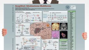 挂图SnapShot: Glioblastoma Multiforme Overview of the key concepts and mechanisms in glioblastoma multiforme biology

 沪公网安备31010102008431号