Belkind-Gerson J et al. (JAN 2013)
Neurogastroenterology and motility : the official journal of the European Gastrointestinal Motility Society 25 1 61--9.e7
Nestin-expressing cells in the gut give rise to enteric neurons and glial cells.
BACKGROUND Neuronal stem cells (NSCs) are promising for neurointestinal disease therapy. Although NSCs have been isolated from intestinal musclularis,their presence in mucosa has not been well described. Mucosa-derived NSCs are accessible endoscopically and could be used autologously. Brain-derived Nestin-positive NSCs are important in endogenous repair and plasticity. The aim was to isolate and characterize mucosa-derived NSCs,determine their relationship to Nestin-expressing cells and to demonstrate their capacity to produce neuroglial networks in vitro and in vivo. METHODS Neurospheres were generated from periventricular brain,colonic muscularis (Musc),and mucosa-submucosa (MSM) of mice expressing green fluorescent protein (GFP) controlled by the Nestin promoter (Nestin-GFP). Neuronal stem cells were also grown as adherent colonies from intestinal mucosal organoids. Their differentiation potential was assessed using immunohistochemistry using glial and neuronal markers. Brain and gut-derived neurospheres were transplanted into explants of chick embryonic aneural hindgut to determine their fate. KEY RESULTS Musc- and MSM-derived neurospheres expressed Nestin and gave rise to cells of neuronal,glial,and mesenchymal lineage. Although Nestin expression in tissue was mostly limited to glia co-labelled with glial fibrillary acid protein (GFAP),neurosphere-derived neurons and glia both expressed Nestin in vitro,suggesting that Nestin+/GFAP+ glial cells may give rise to new neurons. Moreover,following transplantation into aneural colon,brain- and gut-derived NSCs were able to differentiate into neurons. CONCLUSIONS & INFERENCES Nestin-expressing intestinal NSCs cells give rise to neurospheres,differentiate into neuronal,glial,and mesenchymal lineages in vitro,generate neurons in vivo and can be isolated from mucosa. Further studies are needed for exploring their potential for treating neuropathies.
View Publication
产品号#:
05700
05701
05702
05703
05704
05715
产品名:
NeuroCult™ 基础培养基(小鼠和大鼠)
NeuroCult™ 扩增添加物(小鼠和大鼠)
NeuroCult™扩增试剂盒(小鼠和大鼠)
NeuroCult™ 分化添加物(小鼠和大鼠)
NeuroCult™ 分化试剂盒(小鼠和大鼠)
NeuroCult™成年中枢神经系统(CNS)组织酶解试剂盒(小鼠和大鼠)
Ostrakhovitch EA et al. (DEC 2012)
Archives of biochemistry and biophysics 528 1 21--31
Directed differentiation of embryonic P19 cells and neural stem cells into neural lineage on conducting PEDOT-PEG and ITO glass substrates.
Differentiation of pluripotent and lineage restricted stem cells such as neural stem cells (NSCs) was studied on conducting substrates of various nature without perturbation of the genome with exogenous genetic material or chemical stimuli. Primary mouse adult neural stem cells (NSCs) and P19 pluripotent embryonal (P19 EC) carcinoma cells were used. Expression levels of neuronal markers β-III-tubulin and neurofilament were evaluated by immunochemistry and flow cytometry. It was shown that the ability of the substrate to induce differentiation directly correlated with its conductivity. Conducting substrates (conducting oxides or doped pi-conjugated organic polymers) with different morphology,structure,and conductivity mechanisms all promoted differentiation of NSC and P19 cells into neuronal lineage to a similar degree without use of additional factors such as poly-L-ornithine coating or retinoic acid,as verified by their morphology and upregulation of the neuronal markers but not astrocyte marker GFAP. However,substrates with low conductance below ca. 10(-4) S cm(-2) did not show this ability. Morphology of differentiating cells was visualized by atomic force microscopy. NSCs cells increased β-III-tubulin expression by 95% and P19 cells by over 30%. Our results suggest that the substrate conductivity is a key factor governing the cell fate. Differentiation of P19 cells into neuronal lineage on conducting substrates was attributed to downregualtion of Akt signaling pathway and increase in expression of dual oxidase 1 (DUOX 1).
View Publication
Chen W et al. (JUN 2014)
Scientific reports 4 5404
Generation of the SCN1A epilepsy mutation in hiPS cells using the TALEN technique.
Human induced pluripotent stem cells (iPSC) can be used to understand the pathological mechanisms of human disease. These cells are a promising source for cell-replacement therapy. However,such studies require genetically defined conditions. Such genetic manipulations can be performed using the novel Transcription Activator-Like Effector Nucleases (TALENs),which generate site-specific double-strand DNA breaks (DSBs) with high efficiency and precision. Combining the TALEN and iPSC methods,we developed two iPS cell lines by generating the point mutation A5768G in the SCN1A gene,which encodes the voltage-gated sodium channel Nav1.1 α subunit. The engineered iPSC maintained pluripotency and successfully differentiated into neurons with normal functional characteristics. The two cell lines differ exclusively at the epilepsy-susceptibility variant. The ability to robustly introduce disease-causing point mutations in normal hiPS cell lines can be used to generate a human cell model for studying epileptic mechanisms and for drug screening.
View Publication
产品号#:
05850
05857
05870
05875
85850
85857
85870
85875
产品名:
mTeSR™1
mTeSR™1
Vazin T et al. (FEB 2014)
Neurobiology of Disease 62 62--72
Efficient derivation of cortical glutamatergic neurons from human pluripotent stem cells: a model system to study neurotoxicity in Alzheimer's disease.
Alzheimer's disease (AD) is among the most prevalent forms of dementia affecting the aging population,and pharmacological therapies to date have not been successful in preventing disease progression. Future therapeutic efforts may benefit from the development of models that enable basic investigation of early disease pathology. In particular,disease-relevant models based on human pluripotent stem cells (hPSCs) may be promising approaches to assess the impact of neurotoxic agents in AD on specific neuronal populations and thereby facilitate the development of novel interventions to avert early disease mechanisms. We implemented an efficient paradigm to convert hPSCs into enriched populations of cortical glutamatergic neurons emerging from dorsal forebrain neural progenitors,aided by modulating Sonic hedgehog (Shh) signaling. Since AD is generally known to be toxic to glutamatergic circuits,we exposed glutamatergic neurons derived from hESCs to an oligomeric pre-fibrillar forms of Aβ known as globulomers"�
View Publication
I. Canals et al. (SEP 2018)
Nature methods 15 9 693--696
Rapid and efficient induction of functional astrocytes from human pluripotent stem cells.
The derivation of astrocytes from human pluripotent stem cells is currently slow and inefficient. We demonstrate that overexpression of the transcription factors SOX9 and NFIB in human pluripotent stem cells rapidly and efficiently yields homogeneous populations of induced astrocytes. In our study these cells exhibited molecular and functional properties resembling those of adult human astrocytes and were deemed suitable for disease modeling. Our method provides new possibilities for the study of human astrocytes in health and disease.
View Publication
产品号#:
05790
05792
05793
05794
05795
85850
85857
85870
85875
100-0276
100-1130
73802
73804
产品名:
BrainPhys™神经元培养基
BrainPhys™神经元培养基和SM1试剂盒
BrainPhys™ 神经元培养基N2-A和SM1试剂盒
BrainPhys™原代神经元试剂盒
BrainPhys™ hPSC 神经元试剂盒
mTeSR™1
mTeSR™1
mTeSR™ Plus
mTeSR™ Plus
Rho激酶抑制剂IV (Dihydrochloride)
Rho激酶抑制剂IV (Dihydrochloride)
Walker TL et al. (APR 2007)
The Journal of neuroscience : the official journal of the Society for Neuroscience 27 14 3734--42
The doublecortin-expressing population in the developing and adult brain contains multipotential precursors in addition to neuronal-lineage cells.
Doublecortin (DCX) has recently been promulgated as a selective marker of cells committed to the neuronal lineage in both the developing and the adult brain. To explore the potential of DCX-positive (DCX+) cells more stringently,these cells were isolated by flow cytometry from the brains of transgenic mice expressing green fluorescent protein under the control of the DCX promoter in embryonic,early postnatal,and adult animals. It was found that virtually all of the cells (99.9%) expressing high levels of DCX (DCX(high)) in the embryonic brain coexpressed the neuronal marker betaIII-tubulin and that this population contained no stem-like cells as demonstrated by lack of neurosphere formation in vitro. However,the DCX+ population from the early postnatal brain and the adult subventricular zone and hippocampus,which expressed low levels of DCX (DCX(low)),was enriched for neurosphere-forming cells,with only a small subpopulation of these cells coexpressing the neuronal markers betaIII-tubulin or microtubule-associated protein 2. Similarly,the DCX(low) population from embryonic day 14 (E14) brain contained neurosphere-forming cells. Only the postnatal cerebellum and adult olfactory bulb contained some DCX(high) cells,which were shown to be similar to the E14 DCX(high) cells in that they had no stem cell activity. Electrophysiological studies confirmed the heterogeneous nature of DCX+ cells,with some cells displaying characteristics of immature or mature neurons,whereas others showed no neuronal characteristics whatsoever. These results indicate that DCX(high) cells,regardless of location,are restricted to the neuronal lineage or are bone fide neurons,whereas some DCX(low) cells retain their multipotentiality.
View Publication
产品号#:
05700
05701
05702
产品名:
NeuroCult™ 基础培养基(小鼠和大鼠)
NeuroCult™ 扩增添加物(小鼠和大鼠)
NeuroCult™扩增试剂盒(小鼠和大鼠)
Pecho-Vrieseling E et al. (AUG 2014)
Nat Neurosci 17 8 1064--1072
Transneuronal propagation of mutant huntingtin contributes to non-cell autonomous pathology in neurons.
In Huntington's disease (HD),whether transneuronal spreading of mutant huntingtin (mHTT) occurs and its contribution to non-cell autonomous damage in brain networks is largely unknown. We found mHTT spreading in three different neural network models: human neurons integrated in the neural network of organotypic brain slices of HD mouse model,an ex vivo corticostriatal slice model and the corticostriatal pathway in vivo. Transneuronal propagation of mHTT was blocked by two different botulinum neurotoxins,each known for specifically inactivating a single critical component of the synaptic vesicle fusion machinery. Moreover,healthy human neurons in HD mouse model brain slices displayed non-cell autonomous changes in morphological integrity that were more pronounced when these neurons bore mHTT aggregates. Altogether,our findings suggest that transneuronal propagation of mHTT might be an important and underestimated contributor to the pathophysiology of HD.
View Publication


 EasySep™小鼠TIL(CD45)正选试剂盒




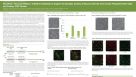
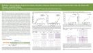 科学海报BrainPhys™ Neuronal Medium Supports the Electrical Activities of Neurons Derived from Human Pluripotent Stem Cells and Primary CNS Tissues in Long-Term Cultures

 沪公网安备31010102008431号