SFN 2016,Advances in Drug Discovery 2017,iForum 2017
Vazin T et al. (FEB 2014)
Neurobiology of Disease 62 62--72
Efficient derivation of cortical glutamatergic neurons from human pluripotent stem cells: a model system to study neurotoxicity in Alzheimer's disease.
Alzheimer's disease (AD) is among the most prevalent forms of dementia affecting the aging population,and pharmacological therapies to date have not been successful in preventing disease progression. Future therapeutic efforts may benefit from the development of models that enable basic investigation of early disease pathology. In particular,disease-relevant models based on human pluripotent stem cells (hPSCs) may be promising approaches to assess the impact of neurotoxic agents in AD on specific neuronal populations and thereby facilitate the development of novel interventions to avert early disease mechanisms. We implemented an efficient paradigm to convert hPSCs into enriched populations of cortical glutamatergic neurons emerging from dorsal forebrain neural progenitors,aided by modulating Sonic hedgehog (Shh) signaling. Since AD is generally known to be toxic to glutamatergic circuits,we exposed glutamatergic neurons derived from hESCs to an oligomeric pre-fibrillar forms of Aβ known as globulomers"�
View Publication
产品号#:
05850
05857
05870
05875
85850
85857
85870
85875
产品名:
mTeSR™1
mTeSR™1
Chen W et al. (JUN 2014)
Scientific reports 4 5404
Generation of the SCN1A epilepsy mutation in hiPS cells using the TALEN technique.
Human induced pluripotent stem cells (iPSC) can be used to understand the pathological mechanisms of human disease. These cells are a promising source for cell-replacement therapy. However,such studies require genetically defined conditions. Such genetic manipulations can be performed using the novel Transcription Activator-Like Effector Nucleases (TALENs),which generate site-specific double-strand DNA breaks (DSBs) with high efficiency and precision. Combining the TALEN and iPSC methods,we developed two iPS cell lines by generating the point mutation A5768G in the SCN1A gene,which encodes the voltage-gated sodium channel Nav1.1 α subunit. The engineered iPSC maintained pluripotency and successfully differentiated into neurons with normal functional characteristics. The two cell lines differ exclusively at the epilepsy-susceptibility variant. The ability to robustly introduce disease-causing point mutations in normal hiPS cell lines can be used to generate a human cell model for studying epileptic mechanisms and for drug screening.
View Publication
产品号#:
05850
05857
05870
05875
85850
85857
85870
85875
产品名:
mTeSR™1
mTeSR™1
Patriarchi T et al. (JUN 2016)
European journal of human genetics : EJHG 24 6 871--880
Imbalance of excitatory/inhibitory synaptic protein expression in iPSC-derived neurons from FOXG1(+/-) patients and in foxg1(+/-) mice.
Rett syndrome (RTT) is a severe neurodevelopmental disorder associated with mutations in either MECP2,CDKL5 or FOXG1. The precise molecular mechanisms that lead to the pathogenesis of RTT have yet to be elucidated. We recently reported that expression of GluD1 (orphan glutamate receptor $\$-1 subunit) is increased in iPSC-derived neurons obtained from patients with mutations in either MECP2 or CDKL5. GluD1 controls synaptic differentiation and shifts the balance between excitatory and inhibitory synapses toward the latter. Thus,an increase in GluD1 might be a critical factor in the etiology of RTT by affecting the excitatory/inhibitory balance in the developing brain. To test this hypothesis,we generated iPSC-derived neurons from FOXG1(+/-) patients. We analyzed mRNA and protein levels of GluD1 together with key markers of excitatory and inhibitory synapses in these iPSC-derived neurons and in Foxg1(+/-) mouse fetal (E11.5) and adult (P70) brains. We found strong correlation between iPSC-derived neurons and fetal mouse brains,where GluD1 and inhibitory synaptic markers (GAD67 and GABA AR-$\$1) were increased,whereas the levels of a number of excitatory synaptic markers (VGLUT1,GluA1,GluN1 and PSD-95) were decreased. In adult mice,GluD1 was decreased along with all GABAergic and glutamatergic markers. Our findings further the understanding of the etiology of RTT by introducing a new pathological event occurring in the brain of FOXG1(+/-) patients during embryonic development and its time-dependent shift toward a general decrease in brain synapses.
View Publication
产品号#:
05850
05857
05870
05875
07923
85850
85857
85870
85875
产品名:
Dispase (1 U/mL)
mTeSR™1
mTeSR™1
Dobie FA and Craig AM (JUL 2011)
The Journal of neuroscience : the official journal of the Society for Neuroscience 31 29 10481--93
Inhibitory synapse dynamics: coordinated presynaptic and postsynaptic mobility and the major contribution of recycled vesicles to new synapse formation.
Dynamics of GABAergic synaptic components have been studied previously over milliseconds to minutes,revealing mobility of postsynaptic scaffolds and receptors. Here we image inhibitory synapses containing fluorescently tagged postsynaptic scaffold Gephyrin,together with presynaptic vesicular GABA transporter (VGAT) or postsynaptic GABA(A) receptor γ2 subunit (GABA(A)Rγ2),over seconds to days in cultured rat hippocampal neurons,revealing modes of inhibitory synapse formation and remodeling. Entire synapses were mobile,translocating rapidly within a confined region and exhibiting greater nonstochastic motion over multihour periods. Presynaptic and postsynaptic components moved in unison,maintaining close apposition while translocating distances of several micrometers. An observed flux in the density of synaptic puncta partially resulted from the apparent merging and splitting of preexisting clusters. De novo formation of inhibitory synapses was observed,marked by the appearance of stably apposed Gephyrin and VGAT clusters at sites previously lacking either component. Coclustering of GABA(A)Rγ2 supports the identification of such new clusters as synapses. Nascent synapse formation occurred by gradual accumulation of components over several hours,with VGAT clustering preceding that of Gephyrin and GABA(A)Rγ2. Comparing VGAT labeling by active uptake of a luminal domain antibody with post hoc immunocytochemistry indicated that recycling vesicles from preexisting boutons significantly contribute to vesicle pools at the majority of new inhibitory synapses. Although new synapses formed primarily on dendrite shafts,some also formed on dendritic protrusions,without apparent interconversion. Altogether,the long-term imaging of GABAergic presynaptic and postsynaptic components reveals complex dynamics and perpetual remodeling with implications for mechanisms of assembly and synaptic integration.
View Publication
产品号#:
05711
100-1281
产品名:
NeuroCult™ SM1 神经添加物
NeuroCult™ SM1 神经添加物
Nekrasov ED et al. (DEC 2016)
Molecular Neurodegeneration 11 1 1--15
Manifestation of Huntington's disease pathology in human induced pluripotent stem cell-derived neurons.
Background: Huntington's disease (HD) is an incurable hereditary neurodegenerative disorder,which manifests itself as a loss of GABAergic medium spiny (GABA MS) neurons in the striatum and caused by an expansion of the CAG repeat in exon 1 of the huntingtin gene. There is no cure for HD,existing pharmaceutical can only relieve its symptoms. Results: Here,induced pluripotent stem cells were established from patients with low CAG repeat expansion in the huntingtin gene,and were then efficiently differentiated into GABA MS-like neurons (GMSLNs) under defined culture conditions. The generated HD GMSLNs recapitulated disease pathology in vitro,as evidenced by mutant huntingtin protein aggregation,increased number of lysosomes/autophagosomes,nuclear indentations,and enhanced neuronal death during cell aging. Moreover,store-operated channel (SOC) currents were detected in the differentiated neurons,and enhanced calcium entry was reproducibly demonstrated in all HD GMSLNs genotypes. Additionally,the quinazoline derivative,EVP4593,reduced the number of lysosomes/autophagosomes and SOC currents in HD GMSLNs and exerted neuroprotective effects during cell aging. Conclusions: Our data is the first to demonstrate the direct link of nuclear morphology and SOC calcium deregulation to mutant huntingtin protein expression in iPSCs-derived neurons with disease-mimetic hallmarks,providing a valuable tool for identification of candidate anti-HD drugs. Our experiments demonstrated that EVP4593 may be a promising anti-HD drug. [ABSTRACT FROM AUTHOR]
View Publication
产品号#:
05854
05855
05850
05857
05870
05875
85850
85857
85870
85875
产品名:
mFreSR™
mFreSR™
mTeSR™1
mTeSR™1
Ito N et al. (APR 2016)
Disease models & mechanisms 9 4 451--462
Decreased N-TAF1 expression in X-linked dystonia-parkinsonism patient-specific neural stem cells.
X-linked dystonia-parkinsonism (XDP) is a hereditary neurodegenerative disorder involving a progressive loss of striatal medium spiny neurons. The mechanisms underlying neurodegeneration are not known,in part because there have been few cellular models available for studying the disease. The XDP haplotype consists of multiple sequence variations in a region of the X chromosome containingTAF1,a large gene with at least 38 exons,and a multiple transcript system (MTS) composed of five unconventional exons. A previous study identified an XDP-specific insertion of a SINE-VNTR-Alu (SVA)-type retrotransposon in intron 32 ofTAF1,as well as a neural-specific TAF1 isoform,N-TAF1,which showed decreased expression in post-mortem XDP brain compared with control tissue. Here,we generated XDP patient and control fibroblasts and induced pluripotent stem cells (iPSCs) in order to further probe cellular defects associated with this disease. As initial validation of the model,we compared expression ofTAF1and MTS transcripts in XDP versus control fibroblasts and iPSC-derived neural stem cells (NSCs). Compared with control cells,XDP fibroblasts exhibited decreased expression ofTAF1transcript fragments derived from exons 32-36,a region spanning the SVA insertion site. N-TAF1,which incorporates an alternative exon (exon 34'),was not expressed in fibroblasts,but was detectable in iPSC-differentiated NSCs at levels that were ∼threefold lower in XDP cells than in controls. These results support the previous findings that N-TAF1 expression is impaired in XDP,but additionally indicate that this aberrant transcription might occur in neural cells at relatively early stages of development that precede neurodegeneration.
View Publication
产品号#:
产品名:
Kim KH et al. (NOV 2015)
PLoS ONE 10 11 e0142693
Transcriptomic analysis of induced pluripotent stem cells derived from patients with bipolar disorder from an old order amish pedigree
Fibroblasts from patients with Type I bipolar disorder (BPD) and their unaffected siblings were obtained from an Old Order Amish pedigree with a high incidence of BPD and reprogrammed to induced pluripotent stem cells (iPSCs). Established iPSCs were subsequently differentiated into neuroprogenitors (NPs) and then to neurons. Transcriptomic microarray analysis was conducted on RNA samples from iPSCs,NPs and neurons matured in culture for either 2 weeks (termed early neurons,E) or 4 weeks (termed late neurons,L). Global RNA profiling indicated that BPD and control iPSCs differentiated into NPs and neurons at a similar rate,enabling studies of differentially expressed genes in neurons from controls and BPD cases. Significant disease-associated differences in gene expression were observed only in L neurons. Specifically,328 genes were differentially expressed between BPD and control L neurons including GAD1,glutamate decarboxylase 1 (2.5 fold) and SCN4B,the voltage gated type IV sodium channel beta subunit (-14.6 fold). Quantitative RT-PCR confirmed the up-regulation of GAD1 in BPD compared to control L neurons. Gene Ontology,GeneGo and Ingenuity Pathway Analysis of differentially regulated genes in L neurons suggest that alterations in RNA biosynthesis and metabolism,protein trafficking as well as receptor signaling pathways may play an important role in the pathophysiology of BPD.
View Publication
产品号#:
05850
05857
05870
05875
85850
85857
85870
85875
27845
27945
27840
27865
27940
27965
产品名:
mTeSR™1
mTeSR™1
Jenkins PM et al. (DEC 2015)
Nanoscale research letters 10 1 972
A nerve guidance conduit with topographical and biochemical cues: potential application using human neural stem cells.
Despite major advances in the pathophysiological understanding of peripheral nerve damage,the treatment of nerve injuries still remains an unmet medical need. Nerve guidance conduits present a promising treatment option by providing a growth-permissive environment that 1) promotes neuronal cell survival and axon growth and 2) directs axonal extension. To this end,we designed an electrospun nerve guidance conduit using a blend of polyurea and poly-caprolactone with both biochemical and topographical cues. Biochemical cues were integrated into the conduit by functionalizing the polyurea with RGD to improve cell attachment. Topographical cues that resemble natural nerve tissue were incorporated by introducing intraluminal microchannels aligned with nanofibers. We determined that electrospinning the polymer solution across a two electrode system with dissolvable sucrose fibers produced a polymer conduit with the appropriate biomimetic properties. Human neural stem cells were cultured on the conduit to evaluate its ability to promote neuronal growth and axonal extension. The nerve guidance conduit was shown to enhance cell survival,migration,and guide neurite extension.
View Publication
产品号#:
05850
05857
05870
05875
85850
85857
85870
85875
产品名:
mTeSR™1
mTeSR™1
Chen C et al. (JUL 2014)
Nature communications 5 4430
Role of astroglia in Down's syndrome revealed by patient-derived human-induced pluripotent stem cells.
Down's syndrome (DS),caused by trisomy of human chromosome 21,is the most common genetic cause of intellectual disability. Here we use induced pluripotent stem cells (iPSCs) derived from DS patients to identify a role for astrocytes in DS pathogenesis. DS astroglia exhibit higher levels of reactive oxygen species and lower levels of synaptogenic molecules. Astrocyte-conditioned medium collected from DS astroglia causes toxicity to neurons,and fails to promote neuronal ion channel maturation and synapse formation. Transplantation studies show that DS astroglia do not promote neurogenesis of endogenous neural stem cells in vivo. We also observed abnormal gene expression profiles from DS astroglia. Finally,we show that the FDA-approved antibiotic drug,minocycline,partially corrects the pathological phenotypes of DS astroglia by specifically modulating the expression of S100B,GFAP,inducible nitric oxide synthase,and thrombospondins 1 and 2 in DS astroglia. Our studies shed light on the pathogenesis and possible treatment of DS by targeting astrocytes with a clinically available drug.
View Publication

 EasySep™小鼠TIL(CD45)正选试剂盒


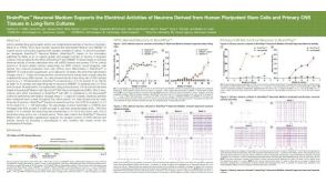 科学海报BrainPhys™ Neuronal Medium Supports the Electrical Activities of Neurons Derived from Human Pluripotent Stem Cells and Primary CNS Tissues in Long-Term Cultures
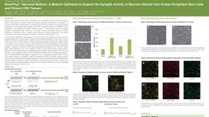

 沪公网安备31010102008431号