Young KM et al. (AUG 2007)
The Journal of neuroscience : the official journal of the Society for Neuroscience 27 31 8286--96
Subventricular zone stem cells are heterogeneous with respect to their embryonic origins and neurogenic fates in the adult olfactory bulb.
We determined the embryonic origins of adult forebrain subventricular zone (SVZ) stem cells by Cre-lox fate mapping in transgenic mice. We found that all parts of the telencephalic neuroepithelium,including the medial ganglionic eminence and lateral ganglionic eminence (LGE) and the cerebral cortex,contribute multipotent,self-renewing stem cells to the adult SVZ. Descendants of the embryonic LGE and cortex settle in ventral and dorsal aspects of the dorsolateral SVZ,respectively. Both populations contribute new (5-bromo-2'-deoxyuridine-labeled) tyrosine hydroxylase- and calretinin-positive interneurons to the adult olfactory bulb. However,calbindin-positive interneurons in the olfactory glomeruli were generated exclusively by LGE-derived stem cells. Thus,different SVZ stem cells have different embryonic origins,colonize different parts of the SVZ,and generate different neuronal progeny,suggesting that some aspects of embryonic patterning are preserved in the adult SVZ. This could have important implications for the design of endogenous stem cell-based therapies in the future.
View Publication
产品号#:
05700
05701
05702
05740
产品名:
NeuroCult™ 基础培养基(小鼠和大鼠)
NeuroCult™ 扩增添加物(小鼠和大鼠)
NeuroCult™扩增试剂盒(小鼠和大鼠)
Gonzalez-Velasquez FJ and Moss MA (JAN 2008)
Journal of neurochemistry 104 2 500--13
Soluble aggregates of the amyloid-beta protein activate endothelial monolayers for adhesion and subsequent transmigration of monocyte cells.
Increasing evidence suggests that the deposition of amyloid plaques,composed primarily of the amyloid-beta protein (Abeta),within the cerebrovasculature is a frequent occurrence in Alzheimer's disease and may play a significant role in disease progression. Accordingly,the pathogenic mechanisms by which Abeta can alter vascular function may have therapeutic implications. Despite observations that Abeta elicits a number of physiological responses in endothelial cells,ranging from alteration of protein expression to cell death,the Abeta species accountable for these responses remains unexplored. In the current study,we show that isolated soluble Abeta aggregation intermediates activate human brain microvascular endothelial cells for both adhesion and subsequent transmigration of monocyte cells in the absence of endothelial cell death and monolayer disruption. In contrast,unaggregated Abeta monomer and mature Abeta fibril fail to induce any change in endothelial adhesion or transmigration. Correlations between average Abeta aggregate size and observed increases in adhesion illustrate that smaller soluble aggregates are more potent activators of endothelium. These results support previous studies demonstrating heightened neuronal activity of soluble Abeta aggregates,including Abeta-derived diffusible ligands,oligomers,and protofibrils,and further show that soluble aggregates also selectively exhibit activity in a vascular cell model.
View Publication
Aberrant Neural Stem Cell Proliferation and Increased Adult Neurogenesis in Mice Lacking Chromatin Protein HMGB2
Neural stem and progenitor cells (NSCs/NPCs) are distinct groups of cells found in the mammalian central nervous system (CNS). Previously we determined that members of the High Mobility Group (HMG) B family of chromatin structural proteins modulate NSC proliferation and self-renewal. Among them HMGB2 was found to be dynamically expressed in proliferating and differentiating NSCs,suggesting that it may regulate NSC maintenance. We report now that Hmgb2(-/-) mice exhibit SVZ hyperproliferation,increased numbers of SVZ NSCs,and a trend towards aberrant increases in newly born neurons in the olfactory bulb (OB) granule cell layer. Increases in the levels of the transcription factor p21 and the Neural cell adhesion molecule (NCAM),along with down-regulation of the transcription/pluripotency factor Oct4 in the Hmgb2-/- SVZ point to a possible pathway for this increased proliferation/differentiation. Our findings suggest that HMGB2 functions as a modulator of neurogenesis in young adult mice through regulation of NSC proliferation,and identify a potential target via which CNS repair could be amplified following trauma or disease-based neuronal degeneration.
View Publication
产品号#:
05700
05701
05702
05715
产品名:
NeuroCult™ 基础培养基(小鼠和大鼠)
NeuroCult™ 扩增添加物(小鼠和大鼠)
NeuroCult™扩增试剂盒(小鼠和大鼠)
NeuroCult™成年中枢神经系统(CNS)组织酶解试剂盒(小鼠和大鼠)
Ji M et al. (SEP 2013)
Science Translational Medicine 5 201 201ra119--201ra119
Rapid, Label-Free Detection of Brain Tumors with Stimulated Raman Scattering Microscopy
Surgery is an essential component in the treatment of brain tumors. However,delineating tumor from normal brain remains a major challenge. We describe the use of stimulated Raman scattering (SRS) microscopy for differentiating healthy human and mouse brain tissue from tumor-infiltrated brain based on histoarchitectural and biochemical differences. Unlike traditional histopathology,SRS is a label-free technique that can be rapidly performed in situ. SRS microscopy was able to differentiate tumor from nonneoplastic tissue in an infiltrative human glioblastoma xenograft mouse model based on their different Raman spectra. We further demonstrated a correlation between SRS and hematoxylin and eosin microscopy for detection of glioma infiltration (κ = 0.98). Finally,we applied SRS microscopy in vivo in mice during surgery to reveal tumor margins that were undetectable under standard operative conditions. By providing rapid intraoperative assessment of brain tissue,SRS microscopy may ultimately improve the safety and accuracy of surgeries where tumor boundaries are visually indistinct.
View Publication
产品号#:
05750
05751
产品名:
NeuroCult™ NS-A 基础培养基(人)
NeuroCult™ NS-A 扩增试剂盒(人)
Baptista S et al. (SEP 2014)
Stem cell research 13 2 329--41
Methamphetamine decreases dentate gyrus stem cell self-renewal and shifts the differentiation towards neuronal fate.
Methamphetamine (METH) is a highly addictive psychostimulant drug of abuse that negatively interferes with neurogenesis. In fact,we have previously shown that METH triggers stem/progenitor cell death and decreases neuronal differentiation in the dentate gyrus (DG). Still,little is known regarding its effect on DG stem cell properties. Herein,we investigate the impact of METH on mice DG stem/progenitor cell self-renewal functions. METH (10nM) decreased DG stem cell self-renewal,while 1nM delayed cell cycle in the G0/G1-to-S phase transition and increased the number of quiescent cells (G0 phase),which correlated with a decrease in cyclin E,pEGFR and pERK1/2 protein levels. Importantly,both drug concentrations (1 or 10nM) did not induce cell death. In accordance with the impairment of self-renewal capacity,METH (10nM) decreased Sox2(+)/Sox2(+) while increased Sox2(-)/Sox2(-) pairs of daughter cells. This effect relied on N-methyl-d-aspartate (NMDA) signaling,which was prevented by the NMDA receptor antagonist,MK-801 (10μM). Moreover,METH (10nM) increased doublecortin (DCX) protein levels consistent with neuronal differentiation. In conclusion,METH alters DG stem cell properties by delaying cell cycle and decreasing self-renewal capacities,mechanisms that may contribute to DG neurogenesis impairment followed by cognitive deficits verified in METH consumers.
View Publication
产品号#:
05707
产品名:
NeuroCult™化学解离试剂盒(小鼠)
Booth L et al. (JUL 2015)
Journal of cellular physiology 230 7 1661--76
GRP78/BiP/HSPA5/Dna K is a universal therapeutic target for human disease.
The chaperone GRP78/Dna K is conserved throughout evolution down to prokaryotes. The GRP78 inhibitor OSU-03012 (AR-12) interacted with sildenafil (Viagra) or tadalafil (Cialis) to rapidly reduce GRP78 levels in eukaryotes and as a single agent reduce Dna K levels in prokaryotes. Similar data with the drug combination were obtained for: HSP70,HSP90,GRP94,GRP58,HSP27,HSP40 and HSP60. OSU-03012/sildenafil treatment killed brain cancer stem cells and decreased the expression of: NPC1 and TIM1; LAMP1; and NTCP1,receptors for Ebola/Marburg/Hepatitis A,Lassa fever,and Hepatitis B viruses,respectively. Pre-treatment with OSU-03012/sildenafil reduced expression of the coxsakie and adenovirus receptor in parallel with it also reducing the ability of a serotype 5 adenovirus or coxsakie virus B4 to infect and to reproduce. Similar data were obtained using Chikungunya,Mumps,Measles,Rubella,RSV,CMV,and Influenza viruses. OSU-03012 as a single agent at clinically relevant concentrations killed laboratory generated antibiotic resistant E. coli and clinical isolate multi-drug resistant N. gonorrhoeae and MRSE which was in bacteria associated with reduced Dna K and Rec A expression. The PDE5 inhibitors sildenafil or tadalafil enhanced OSU-03012 killing in N. gonorrhoeae and MRSE and low marginally toxic doses of OSU-03012 could restore bacterial sensitivity in N. gonorrhoeae to multiple antibiotics. Thus,Dna K and bacterial phosphodiesterases are novel antibiotic targets,and inhibition of GRP78 is of therapeutic utility for cancer and also for bacterial and viral infections.
View Publication
产品号#:
05750
05751
产品名:
NeuroCult™ NS-A 基础培养基(人)
NeuroCult™ NS-A 扩增试剂盒(人)
Binder ZA et al. ( 2016)
PloS one 11 3 e0150271
Establishment and Biological Characterization of a Panel of Glioblastoma Multiforme (GBM) and GBM Variant Oncosphere Cell Lines.
OBJECTIVE Human tumor cell lines form the basis of the majority of present day laboratory cancer research. These models are vital to studying the molecular biology of tumors and preclinical testing of new therapies. When compared to traditional adherent cell lines,suspension cell lines recapitulate the genetic profiles and histologic features of glioblastoma multiforme (GBM) with higher fidelity. Using a modified neural stem cell culture technique,here we report the characterization of GBM cell lines including GBM variants. METHODS Tumor tissue samples were obtained intra-operatively and cultured in neural stem cell conditions containing growth factors. Tumor lines were characterized in vitro using differentiation assays followed by immunostaining for lineage-specific markers. In vivo tumor formation was assayed by orthotopic injection in nude mice. Genetic uniqueness was confirmed via short tandem repeat (STR) DNA profiling. RESULTS Thirteen oncosphere lines derived from GBM and GBM variants,including a GBM with PNET features and a GBM with oligodendroglioma component,were established. All unique lines showed distinct genetic profiles by STR profiling. The lines assayed demonstrated a range of in vitro growth rates. Multipotency was confirmed using in vitro differentiation. Tumor formation demonstrated histologic features consistent with high grade gliomas,including invasion,necrosis,abnormal vascularization,and high mitotic rate. Xenografts derived from the GBM variants maintained histopathological features of the primary tumors. CONCLUSIONS We have generated and characterized GBM suspension lines derived from patients with GBMs and GBM variants. These oncosphere cell lines will expand the resources available for preclinical study.
View Publication
产品号#:
05750
05751
产品名:
NeuroCult™ NS-A 基础培养基(人)
NeuroCult™ NS-A 扩增试剂盒(人)
Biasini E et al. (FEB 2013)
Journal of Neuroscience 33 6 2408--2418
A Mutant Prion Protein Sensitizes Neurons to Glutamate-Induced Excitotoxicity
Growing evidence suggests that a physiological activity of the cellular prion protein (PrP(C)) plays a crucial role in several neurodegenerative disorders,including prion and Alzheimer's diseases. However,how the functional activity of PrP(C) is subverted to deliver neurotoxic signals remains uncertain. Transgenic (Tg) mice expressing PrP with a deletion of residues 105-125 in the central region (referred to as ΔCR PrP) provide important insights into this problem. Tg(ΔCR) mice exhibit neonatal lethality and massive degeneration of cerebellar granule neurons,a phenotype that is dose dependently suppressed by the presence of wild-type PrP. When expressed in cultured cells,ΔCR PrP induces large,ionic currents that can be detected by patch-clamping techniques. Here,we tested the hypothesis that abnormal ion channel activity underlies the neuronal death seen in Tg(ΔCR) mice. We find that ΔCR PrP induces abnormal ionic currents in neurons in culture and in cerebellar slices and that this activity sensitizes the neurons to glutamate-induced,calcium-mediated death. In combination with ultrastructural and biochemical analyses,these results demonstrate a role for glutamate-induced excitotoxicity in PrP-mediated neurodegeneration. A similar mechanism may operate in other neurodegenerative disorders attributable to toxic,β-rich oligomers that bind to PrP(C).
View Publication
产品号#:
05700
05702
05704
产品名:
NeuroCult™ 基础培养基(小鼠和大鼠)
NeuroCult™扩增试剂盒(小鼠和大鼠)
NeuroCult™ 分化试剂盒(小鼠和大鼠)
Yamazaki K et al. (DEC 2016)
Journal of Biomolecular Screening 21 10 1054--1064
Functional Comparison of Neuronal Cells Differentiated from Human Induced Pluripotent Stem CellDerived Neural Stem Cells under Different Oxygen and Medium Conditions
Because neurons are difficult to obtain from humans,generating functional neurons from human induced pluripotent stem cells (hiPSCs) is important for establishing physiological or disease-relevant screening systems for drug discovery. To examine the culture conditions leading to efficient differentiation of functional neural cells,we investigated the effects of oxygen stress (2% or 20% O2) and differentiation medium (DMEM/F12:Neurobasal-based [DN] or commercial [PhoenixSongs Biologicals; PS]) on the expression of genes related to neural differentiation,glutamate receptor function,and the formation of networks of neurons differentiated from hiPSCs (201B7) via long-term self-renewing neuroepithelial-like stem (lt-NES) cells. Expression of genes related to neural differentiation occurred more quickly in PS and/or 2% O2 than in DN and/or 20% O2,resulting in high responsiveness of neural cells to glutamate,N-methyl-d-aspartate (NMDA),α-amino-3-hydroxy-5-methyl-4-isoxazolepropionate (AMPA),and (S)-3,5-d...
View Publication
产品号#:
05832
产品名:
STEMdiff™ 神经花环选择试剂
Yamamizu K et al. (DEC 2013)
Stem Cell Reports 1 6 545--559
Identification of Transcription Factors for Lineage-Specific ESC Differentiation
A network of transcription factors (TFs) determines cell identity,but identity can be altered by overexpressing a combination of TFs. However,choosing and verifying combinations of TFs for specific cell differentiation have been daunting due to the large number of possible combinations of 2,000 TFs. Here,we report the identification of individual TFs for lineage-specific cell differentiation based on the correlation matrix of global gene expression profiles. The overexpression of identified TFs-Myod1,Mef2c,Esx1,Foxa1,Hnf4a,Gata2,Gata3,Myc,Elf5,Irf2,Elf1,Sfpi1,Ets1,Smad7,Nr2f1,Sox11,Dmrt1,Sox9,Foxg1,Sox2,or Ascl1-can direct efficient,specific,and rapid differentiation into myocytes,hepatocytes,blood cells,and neurons. Furthermore,transfection of synthetic mRNAs of TFs generates their appropriate target cells. These results demonstrate both the utility of this approach to identify potent TFs for cell differentiation,and the unanticipated capacity of single TFs directly guides differentiation to specific lineage fates.
View Publication

 EasySep™小鼠TIL(CD45)正选试剂盒


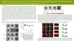 科学海报STEMdiff™ Cerebral Organoid Kit: A New Tool for the Culture of 3D Brain Organoids Derived from hPSCs
 沪公网安备31010102008431号