Pecho-Vrieseling E et al. (AUG 2014)
Nat Neurosci 17 8 1064--1072
Transneuronal propagation of mutant huntingtin contributes to non-cell autonomous pathology in neurons.
In Huntington's disease (HD),whether transneuronal spreading of mutant huntingtin (mHTT) occurs and its contribution to non-cell autonomous damage in brain networks is largely unknown. We found mHTT spreading in three different neural network models: human neurons integrated in the neural network of organotypic brain slices of HD mouse model,an ex vivo corticostriatal slice model and the corticostriatal pathway in vivo. Transneuronal propagation of mHTT was blocked by two different botulinum neurotoxins,each known for specifically inactivating a single critical component of the synaptic vesicle fusion machinery. Moreover,healthy human neurons in HD mouse model brain slices displayed non-cell autonomous changes in morphological integrity that were more pronounced when these neurons bore mHTT aggregates. Altogether,our findings suggest that transneuronal propagation of mHTT might be an important and underestimated contributor to the pathophysiology of HD.
View Publication
产品号#:
05850
05857
05870
05875
85850
85857
85870
85875
产品名:
mTeSR™1
mTeSR™1
Calabrese B and Halpain S (DEC 2014)
Neuroreport 25 17 1331--7
Lithium prevents aberrant NMDA-induced F-actin reorganization in neurons.
Increasing evidence suggests that cellular stress may underlie mood disorders such as bipolar disorder and major depression,particularly as lithium and its targets can protect against neuronal cell death. Here we describe N-methyl-D-aspartate (NMDA)-induced filamentous actin reorganization (NIFAR) as a useful in-vitro model for studying acute neurocellular stress and investigating the effects of mood stabilizers. Brief incubation of cultured neurons with NMDA (50 µM,5 min) induces marked reorganization of F-actin within the somatodendritic domain of a majority of neurons. During NIFAR,F-actin is rapidly depleted from dendritic spines and aberrantly aggregates within the dendrite shaft. The widely used mood stabilizer lithium chloride prevented NIFAR in a time-dependent and dose-dependent manner,consistent with its known efficacy in treating bipolar disorder. Inhibitors of the lithium target glycogen synthase kinase 3 and its upstream activator phosphoinositide-3-kinase also prevented NIFAR. The antidepressant compounds imipramine and fluoxetine also attenuated NIFAR. These findings have potential relevance to neuropsychiatric diseases characterized by excessive glutamate receptor activity and synaptotoxicity. We propose that protection of the dendritic actin cytoskeleton may be a common mechanism shared by various mood stabilizers.
View Publication
产品号#:
05711
100-1281
产品名:
NeuroCult™ SM1 神经添加物
NeuroCult™ SM1 神经添加物
Calabrese B et al. (APR 2014)
PLoS ONE 9 4 e94787
Activity-Dependent Dendritic Spine Shrinkage and Growth Involve Downregulation of Cofilin via Distinct Mechanisms
A current model posits that cofilin-dependent actin severing negatively impacts dendritic spine volume. Studies suggested that increased cofilin activity underlies activity-dependent spine shrinkage,and that reduced cofilin activity induces activity-dependent spine growth. We suggest instead that both types of structural plasticity correlate with decreased cofilin activity. However,the mechanism of inhibition determines the outcome for spine morphology. RNAi in rat hippocampal cultures demonstrates that cofilin is essential for normal spine maintenance. Cofilin-F-actin binding and filament barbed-end production decrease during the early phase of activity-dependent spine shrinkage; cofilin concentration also decreases. Inhibition of the cathepsin B/L family of proteases prevents both cofilin loss and spine shrinkage. Conversely,during activity-dependent spine growth,LIM kinase stimulates cofilin phosphorylation,which activates phospholipase D-1 to promote actin polymerization. These results implicate novel molecular mechanisms and prompt a revision of the current model for how cofilin functions in activity-dependent structural plasticity.
View Publication
Gupta S et al. (DEC 2017)
Journal of Neurochemistry
Fibroblast growth factor 2 regulates activity and gene expression of human post-mitotic excitatory neurons
Many neuropsychiatric disorders are thought to result from subtle changes in neural circuit formation. We used human embryonic stem cells and induced pluripotent stem cells (hiPSCs) to model mature,post-mitotic excitatory neurons and examine effects of fibroblast growth factor 2 (FGF2). FGF2 gene expression is known to be altered in brain regions of major depressive disorder (MDD) patients and FGF2 has anti-depressive effects in animal models of depression. We generated stable inducible neurons (siNeurons) conditionally expressing human neurogenin-2 (NEUROG2) to generate a homogenous population of post-mitotic excitatory neurons and study the functional as well as the transcriptional effects of FGF2. Upon induction of NEUROG2 with doxycycline,the vast majority of cells are post-mitotic,and the gene expression profile recapitulates that of excitatory neurons within 6 days. Using hES cell lines that inducibly express NEUROG2 as well as GCaMP6f,we were able to characterize spontaneous calcium activity in these neurons and show that calcium transients increase in the presence of FGF2. The FGF2-responsive genes were determined by RNA-Seq. FGF2-regulated genes previously identified in non-neuronal cell types were up-regulated (EGR1,ETV4,SPRY4,and DUSP6) as a result of chronic FGF2 treatment of siNeurons. Novel neuron-specific genes were also identified that may mediate FGF2-dependent increases in synaptic efficacy including NRXN3,SYT2,and GALR1. Since several of these genes have been implicated in MDD previously,these results will provide the basis for more mechanistic studies of the role of FGF2 in MDD.
View Publication
产品号#:
05790
05792
05793
05794
05795
85850
85857
85870
85875
05835
05839
产品名:
BrainPhys™神经元培养基
BrainPhys™神经元培养基和SM1试剂盒
BrainPhys™ 神经元培养基N2-A和SM1试剂盒
BrainPhys™原代神经元试剂盒
BrainPhys™ hPSC 神经元试剂盒
mTeSR™1
mTeSR™1
STEMdiff™ 神经诱导培养基
STEMdiff™ 神经诱导培养基
Dobie FA and Craig AM (JUL 2011)
The Journal of neuroscience : the official journal of the Society for Neuroscience 31 29 10481--93
Inhibitory synapse dynamics: coordinated presynaptic and postsynaptic mobility and the major contribution of recycled vesicles to new synapse formation.
Dynamics of GABAergic synaptic components have been studied previously over milliseconds to minutes,revealing mobility of postsynaptic scaffolds and receptors. Here we image inhibitory synapses containing fluorescently tagged postsynaptic scaffold Gephyrin,together with presynaptic vesicular GABA transporter (VGAT) or postsynaptic GABA(A) receptor γ2 subunit (GABA(A)Rγ2),over seconds to days in cultured rat hippocampal neurons,revealing modes of inhibitory synapse formation and remodeling. Entire synapses were mobile,translocating rapidly within a confined region and exhibiting greater nonstochastic motion over multihour periods. Presynaptic and postsynaptic components moved in unison,maintaining close apposition while translocating distances of several micrometers. An observed flux in the density of synaptic puncta partially resulted from the apparent merging and splitting of preexisting clusters. De novo formation of inhibitory synapses was observed,marked by the appearance of stably apposed Gephyrin and VGAT clusters at sites previously lacking either component. Coclustering of GABA(A)Rγ2 supports the identification of such new clusters as synapses. Nascent synapse formation occurred by gradual accumulation of components over several hours,with VGAT clustering preceding that of Gephyrin and GABA(A)Rγ2. Comparing VGAT labeling by active uptake of a luminal domain antibody with post hoc immunocytochemistry indicated that recycling vesicles from preexisting boutons significantly contribute to vesicle pools at the majority of new inhibitory synapses. Although new synapses formed primarily on dendrite shafts,some also formed on dendritic protrusions,without apparent interconversion. Altogether,the long-term imaging of GABAergic presynaptic and postsynaptic components reveals complex dynamics and perpetual remodeling with implications for mechanisms of assembly and synaptic integration.
View Publication
产品号#:
05711
100-1281
产品名:
NeuroCult™ SM1 神经添加物
NeuroCult™ SM1 神经添加物
Brigidi GS et al. (SEP 2015)
Nature communications 6 8200
Activity-regulated trafficking of the palmitoyl-acyl transferase DHHC5.
Synaptic plasticity is mediated by the dynamic localization of proteins to and from synapses. This is controlled,in part,through activity-induced palmitoylation of synaptic proteins. Here we report that the ability of the palmitoyl-acyl transferase,DHHC5,to palmitoylate substrates in an activity-dependent manner is dependent on changes in its subcellular localization. Under basal conditions,DHHC5 is bound to PSD-95 and Fyn kinase,and is stabilized at the synaptic membrane through Fyn-mediated phosphorylation of a tyrosine residue within the endocytic motif of DHHC5. In contrast,DHHC5's substrate,δ-catenin,is highly localized to dendritic shafts,resulting in the segregation of the enzyme/substrate pair. Neuronal activity disrupts DHHC5/PSD-95/Fyn kinase complexes,enhancing DHHC5 endocytosis,its translocation to dendritic shafts and its association with δ-catenin. Following DHHC5-mediated palmitoylation of δ-catenin,DHHC5 and δ-catenin are trafficked together back into spines where δ-catenin increases cadherin stabilization and recruitment of AMPA receptors to the synaptic membrane.
View Publication
产品号#:
05711
100-1281
产品名:
NeuroCult™ SM1 神经添加物
NeuroCult™ SM1 神经添加物
Chen C et al. (JUL 2014)
Nature communications 5 4430
Role of astroglia in Down's syndrome revealed by patient-derived human-induced pluripotent stem cells.
Down's syndrome (DS),caused by trisomy of human chromosome 21,is the most common genetic cause of intellectual disability. Here we use induced pluripotent stem cells (iPSCs) derived from DS patients to identify a role for astrocytes in DS pathogenesis. DS astroglia exhibit higher levels of reactive oxygen species and lower levels of synaptogenic molecules. Astrocyte-conditioned medium collected from DS astroglia causes toxicity to neurons,and fails to promote neuronal ion channel maturation and synapse formation. Transplantation studies show that DS astroglia do not promote neurogenesis of endogenous neural stem cells in vivo. We also observed abnormal gene expression profiles from DS astroglia. Finally,we show that the FDA-approved antibiotic drug,minocycline,partially corrects the pathological phenotypes of DS astroglia by specifically modulating the expression of S100B,GFAP,inducible nitric oxide synthase,and thrombospondins 1 and 2 in DS astroglia. Our studies shed light on the pathogenesis and possible treatment of DS by targeting astrocytes with a clinically available drug.
View Publication
产品号#:
05850
05857
05870
05875
85850
85857
85870
85875
产品名:
mTeSR™1
mTeSR™1
Katori S et al. (JUL 2009)
The Journal of neuroscience : the official journal of the Society for Neuroscience 29 29 9137--47
Protocadherin-alpha family is required for serotonergic projections to appropriately innervate target brain areas.
Serotonergic axons from the raphe nuclei in the brainstem project to every region of the brain,where they make connections through their extensive terminal arborizations. This serotonergic innervation contributes to various normal behaviors and psychiatric disorders. The protocadherin-alpha (Pcdha) family of clustered protocadherins consists of 14 cadherin-related molecules generated from a single gene cluster. We found that the Pcdhas were strongly expressed in the serotonergic neurons. To elucidate their roles,we examined serotonergic fibers in a mouse mutant (Pcdha(Delta CR/Delta CR)) lacking the Pcdha cytoplasmic region-encoding exons,which are common to the gene cluster. In the first week after birth,the distribution pattern of serotonergic fibers in Pcdha(Delta CR/Delta CR) mice was similar to wild-type,but by 3 weeks of age,when the serotonergic axonal termini complete their arborizations,the distribution of the projections was abnormal. In some target regions,notably the globus pallidus and substantia nigra,the normally even distribution of serotonin axonal terminals was,in the mutants,dense at the periphery of each region,but sparse in the center. In the stratum lacunosum-molecular of the hippocampus,the mutants showed denser serotonergic innervation than in wild-type,and in the dentate gyrus of the hippocampus and the caudate-putamen,the innervation was sparser. Together,the abnormalities suggested that Pcdha proteins are important in the late-stage maturation of serotonergic projections. Further examination of alternatively spliced exons encoding the cytoplasmic tail showed that the A-type (but not the B-type) cytoplasmic tail was essential for the normal development of serotonergic projections.
View Publication
产品号#:
03800
03801
03802
03803
03804
03805
03806
产品名:
ClonaCell™-HY杂交瘤试剂盒
ClonaCell™-HY培养基A
ClonaCell™-HY 培养基 B
ClonaCell™-HY 培养基 C
ClonaCell™-HY 培养基 D
ClonaCell™-HY 培养基 E
ClonaCell™-HY PEG
A. M. Tukker et al. (JUL 2018)
Neurotoxicology 67 215--225
Human iPSC-derived neuronal models for in vitro neurotoxicity assessment.
Neurotoxicity testing still relies on ethically debated,expensive and time consuming in vivo experiments,which are unsuitable for high-throughput toxicity screening. There is thus a clear need for a rapid in vitro screening strategy that is preferably based on human-derived neurons to circumvent interspecies translation. Recent availability of commercially obtainable human induced pluripotent stem cell (hiPSC)-derived neurons and astrocytes holds great promise in assisting the transition from the current standard of rat primary cortical cultures to an animal-free alternative. We therefore composed several hiPSC-derived neuronal models with different ratios of excitatory and inhibitory neurons in the presence or absence of astrocytes. Using immunofluorescent stainings and multi-well micro-electrode array (mwMEA) recordings we demonstrate that these models form functional neuronal networks that become spontaneously active. The differences in development of spontaneous neuronal activity and bursting behavior as well as spiking patterns between our models confirm the importance of the presence of astrocytes. Preliminary neurotoxicity assessment demonstrates that these cultures can be modulated with known seizurogenic compounds,such as picrotoxin (PTX) and endosulfan,and the neurotoxicant methylmercury (MeHg). However,the chemical-induced effects on different parameters for neuronal activity,such as mean spike rate (MSR) and mean burst rate (MBR),may depend on the ratio of inhibitory and excitatory neurons. Our results thus indicate that hiPSC-derived neuronal models must be carefully designed and characterized prior to large-scale use in neurotoxicity screening.
View Publication
产品号#:
05790
05792
05793
05794
05795
R1061
R1034
R1116
产品名:
BrainPhys™神经元培养基
BrainPhys™神经元培养基和SM1试剂盒
BrainPhys™ 神经元培养基N2-A和SM1试剂盒
BrainPhys™原代神经元试剂盒
BrainPhys™ hPSC 神经元试剂盒
M. van den Hurk et al. ( 2018)
Frontiers in Molecular Neuroscience
Patch-Seq Protocol to Analyze the Electrophysiology, Morphology and Transcriptome of Whole Single Neurons Derived From Human Pluripotent Stem Cells
The human brain is composed of a complex assembly of about 171 billion heterogeneous cellular units (86 billion neurons and 85 billion non-neuronal glia cells). A comprehensive description of brain cells is necessary to understand the nervous system in health and disease. Recently,advances in genomics have permitted the accurate analysis of the full transcriptome of single cells (scRNA-seq). We have built upon such technical progress to combine scRNA-seq with patch-clamping electrophysiological recording and morphological analysis of single human neurons in vitro. This new powerful method,referred to as Patch-seq,enables a thorough,multimodal profiling of neurons and permits us to expose the links between functional properties,morphology,and gene expression. Here,we present a detailed Patch-seq protocol for isolating single neurons from in vitro neuronal cultures. We have validated the Patch-seq whole-transcriptome profiling method with human neurons generated from embryonic and induced pluripotent stem cells (ESCs/iPSCs) derived from healthy subjects,but the procedure may be applied to any kind of cell type in vitro. Patch-seq may be used on neurons in vitro to profile cell types and states in depth to unravel the human molecular basis of neuronal diversity and investigate the cellular mechanisms underlying brain disorders.
View Publication

 EasySep™小鼠TIL(CD45)正选试剂盒


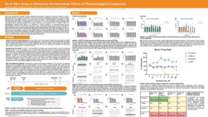 科学海报An In Vitro Assay to Determine the Neurotoxic Effects of Pharmacological Compounds
 沪公网安备31010102008431号