Bagó et al. (FEB 2017)
Science Translational Medicine 9 375 eaah6510
Tumor-homing cytotoxic human induced neural stem cells for cancer therapy
Engineered neural stem cells (NSCs) are a promising approach to treating glioblastoma (GBM). The ideal NSC drug carrier for clinical use should be easily isolated and autologous to avoid immune rejection. We transdifferentiated (TD) human fibroblasts into tumor-homing early-stage induced NSCs (h-iNSC(TE)),engineered them to express optical reporters and different therapeutic gene products,and assessed the tumor-homing migration and therapeutic efficacy of cytotoxic h-iNSC(TE) in patient-derived GBM models of surgical and nonsurgical disease. Molecular and functional analysis revealed that our single-factor SOX2 TD strategy converted human skin fibroblasts into h-iNSC(TE) that were nestin(+) and expressed pathways associated with tumor-homing migration in 4 days. Time-lapse motion analysis showed that h-iNSC(TE) rapidly migrated to human GBM cells and penetrated human GBM spheroids,a process inhibited by blockade of CXCR4. Serial imaging showed that h-iNSC(TE) delivery of the proapoptotic agent tumor necrosis factor-α-related apoptosis-inducing ligand (TRAIL) reduced the size of solid human GBM xenografts 250-fold in 3 weeks and prolonged median survival from 22 to 49 days. Additionally,h-iNSC(TE) thymidine kinase/ganciclovir enzyme/prodrug therapy (h-iNSC(TE)-TK) reduced the size of patient-derived GBM xenografts 20-fold and extended survival from 32 to 62 days. Mimicking clinical NSC therapy,h-iNSC(TE)-TK therapy delivered into the postoperative surgical resection cavity delayed the regrowth of residual GBMs threefold and prolonged survival from 46 to 60 days. These results suggest that TD of human skin into h-iNSC(TE) is a platform for creating tumor-homing cytotoxic cell therapies for cancer,where the potential to avoid carrier rejection could maximize treatment durability in human trials.
View Publication
Although human induced pluripotent stem cells (hiPSCs) hold great potential for the study of human diseases affecting disparate cell types,they have been underutilized in seeking mechanistic insights into the pathogenesis of congenital craniofacial disorders. Craniofrontonasal syndrome (CFNS) is a rare X-linked disorder caused by mutations in EFNB1 and characterized by craniofacial,skeletal,and neurological anomalies. Heterozygous females are more severely affected than hemizygous males,a phenomenon termed cellular interference that involves mosaicism for EPHRIN-B1 function. Although the mechanistic basis for cellular interference in CFNS has been hypothesized to involve Eph/ephrin-mediated cell segregation,no direct evidence for this has been demonstrated. Here,by generating hiPSCs from CFNS patients,we demonstrate that mosaicism for EPHRIN-B1 expression induced by random X inactivation in heterozygous females results in robust cell segregation in human neuroepithelial cells,thus supplying experimental evidence that Eph/ephrin-mediated cell segregation is relevant to pathogenesis in human CFNS patients.
View Publication
产品号#:
05835
05839
08581
08582
产品名:
STEMdiff™ 神经诱导培养基
STEMdiff™ 神经诱导培养基
STEMdiff™SMADi神经诱导试剂盒
STEMdiff™SMADi神经诱导试剂盒,2套
Keller GM (DEC 1995)
Current opinion in cell biology 7 6 862--9
In vitro differentiation of embryonic stem cells.
Under appropriate conditions in culture,embryonic stem cells will differentiate and form embryoid bodies that have been shown to contain cells of the hematopoietic,endothelial,muscle and neuronal lineages. Many aspects of the lineage-specific differentiation programs observed within the embryoid bodies reflect those found in the embryo,indicating that this model system provides access to early cell populations that develop in a normal fashion. Recent studies involving the differentiation of genetically altered embryonic stem cells highlight the potential of this in vitro differentiation system for defining the function of genes in early development.
View Publication
Jackson TC et al. (FEB 2018)
Experimental Neurology 300 232--246
BrainPhys increases neurofilament levels in CNS cultures, and facilitates investigation of axonal damage after a mechanical stretch-injury in vitro
Neurobasal®/B27 is a gold standard culture media used to study primary neurons in vitro. An alternative media (BrainPhys®/SM1) was recently developed which robustly enhances neuronal activity vs. Neurobasal® or DMEM. To the best of our knowledge BrainPhys® has not been explored in the setting of neuronal injury. Here we characterized the utility of BrainPhys® in a model of in vitro mechanical-stretch injury. METHODS/RESULTSPrimary rat cortical neurons were maintained in classic Neurobasal®,or sequentially maintained in Neurocult® followed by BrainPhys® (hereafter simply referred to as BrainPhys® maintained neurons?). The levels of axonal markers and proteins involved in neurotransmission were compared on day in vitro 10 (DIV10). BrainPhys® maintained neurons had higher levels of GluN2B,GluR1,Neurofilament light/heavy chain (NF-L & NF-H),and protein phosphatase 2 A (PP2A) vs. neurons in Neurobasal®. Mechanical stretch-injury (50ms/54% biaxial stretch) to BrainPhys® maintained neurons modestly (albeit significantly) increased 24h lactate dehydrogenase (LDH) levels but markedly decreased axonal NF-L levels post-injury vs. uninjured controls or neurons given a milder 38% stretch-injury. Furthermore,two 54% stretch-injuries (in tandem) exacerbated 24h LDH release,increased α-spectrin breakdown products (SBDPs),and decreased Tau levels. Also,BrainPhys® maintained cultures had decreased markers of cell damage 24h after a single 54% stretch-injury vs. neurons in Neurobasal®. Finally,we tested the hypothesis that lentivirus mediated overexpression of the pro-death protein RBM5 exacerbates neuronal and/or axonal injury in primary CNS cultures. RBM5 overexpression vs. empty-vector controls increased 24h LDH release,and SBDP levels,after a single 54% stretch-injury but did not affect NF-L levels or Tau. CONCLUSIONBrainPhys® is a promising new reagent which facilities the investigation of molecular targets involved in axonal and/or neuronal injury in vitro.
View Publication
产品号#:
05790
05792
05793
05794
05795
产品名:
BrainPhys™神经元培养基
BrainPhys™神经元培养基和SM1试剂盒
BrainPhys™ 神经元培养基N2-A和SM1试剂盒
BrainPhys™原代神经元试剂盒
BrainPhys™ hPSC 神经元试剂盒
Kayama T et al. (JAN 2018)
Biochemical and Biophysical Research Communications 495 1 1028--1033
Temporally coordinated spiking activity of human induced pluripotent stem cell-derived neurons co-cultured with astrocytes
In culture conditions,human induced-pluripotent stem cells (hiPSC)-derived neurons form synaptic connections with other cells and establish neuronal networks,which are expected to be an in vitro model system for drug discovery screening and toxicity testing. While early studies demonstrated effects of co-culture of hiPSC-derived neurons with astroglial cells on survival and maturation of hiPSC-derived neurons,the population spiking patterns of such hiPSC-derived neurons have not been fully characterized. In this study,we analyzed temporal spiking patterns of hiPSC-derived neurons recorded by a multi-electrode array system. We discovered that specific sets of hiPSC-derived neurons co-cultured with astrocytes showed more frequent and highly coherent non-random synchronized spike trains and more dynamic changes in overall spike patterns over time. These temporally coordinated spiking patterns are physiological signs of organized circuits of hiPSC-derived neurons and suggest benefits of co-culture of hiPSC-derived neurons with astrocytes.
View Publication
On-demand optogenetic activation of human stem-cell-derived neurons
The widespread application of human stem-cell-derived neurons for functional studies is impeded by complicated differentiation protocols,immaturity,and deficient optogene expression as stem cells frequently lose transgene expression over time. Here we report a simple but precise Cre-loxP-based strategy for generating conditional,and thereby stable,optogenetic human stem-cell lines. These cells can be easily and efficiently differentiated into functional neurons,and optogene expression can be triggered by administering Cre protein to the cultures. This conditional expression system may be applied to stem-cell-derived neurons whenever timed transgene expression could help to overcome silencing at the stem-cell level.
View Publication
产品号#:
05711
05790
05792
05793
05794
05795
100-1281
产品名:
NeuroCult™ SM1 神经添加物
BrainPhys™神经元培养基
BrainPhys™神经元培养基和SM1试剂盒
BrainPhys™ 神经元培养基N2-A和SM1试剂盒
BrainPhys™原代神经元试剂盒
BrainPhys™ hPSC 神经元试剂盒
NeuroCult™ SM1 神经添加物
Gupta S et al. (DEC 2017)
Journal of Neurochemistry
Fibroblast growth factor 2 regulates activity and gene expression of human post-mitotic excitatory neurons
Many neuropsychiatric disorders are thought to result from subtle changes in neural circuit formation. We used human embryonic stem cells and induced pluripotent stem cells (hiPSCs) to model mature,post-mitotic excitatory neurons and examine effects of fibroblast growth factor 2 (FGF2). FGF2 gene expression is known to be altered in brain regions of major depressive disorder (MDD) patients and FGF2 has anti-depressive effects in animal models of depression. We generated stable inducible neurons (siNeurons) conditionally expressing human neurogenin-2 (NEUROG2) to generate a homogenous population of post-mitotic excitatory neurons and study the functional as well as the transcriptional effects of FGF2. Upon induction of NEUROG2 with doxycycline,the vast majority of cells are post-mitotic,and the gene expression profile recapitulates that of excitatory neurons within 6 days. Using hES cell lines that inducibly express NEUROG2 as well as GCaMP6f,we were able to characterize spontaneous calcium activity in these neurons and show that calcium transients increase in the presence of FGF2. The FGF2-responsive genes were determined by RNA-Seq. FGF2-regulated genes previously identified in non-neuronal cell types were up-regulated (EGR1,ETV4,SPRY4,and DUSP6) as a result of chronic FGF2 treatment of siNeurons. Novel neuron-specific genes were also identified that may mediate FGF2-dependent increases in synaptic efficacy including NRXN3,SYT2,and GALR1. Since several of these genes have been implicated in MDD previously,these results will provide the basis for more mechanistic studies of the role of FGF2 in MDD.
View Publication

 EasySep™小鼠TIL(CD45)正选试剂盒


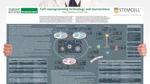 挂图Cell-Reprogramming Technology and Neuroscience Details on human iPSC-derived models of neuropsychiatric and neurodegenerative disorders
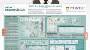 挂图Building Three-Dimensional Human Brain Organoids Overview of brain organogenesis and the applications of brain organoids in studying the development and maturation of the nervous system

 沪公网安备31010102008431号