Kishigami S et al. (FEB 2006)
Biochemical and biophysical research communications 340 1 183--9
Significant improvement of mouse cloning technique by treatment with trichostatin A after somatic nuclear transfer.
The low success rate of animal cloning by somatic cell nuclear transfer (SCNT) is believed to be associated with epigenetic errors including abnormal DNA hypermethylation. Recently,we elucidated by using round spermatids that,after nuclear transfer,treatment of zygotes with trichostatin A (TSA),an inhibitor of histone deacetylase,can remarkably reduce abnormal DNA hypermethylation depending on the origins of transferred nuclei and their genomic regions [S. Kishigami,N. Van Thuan,T. Hikichi,H. Ohta,S. Wakayama. E. Mizutani,T. Wakayama,Epigenetic abnormalities of the mouse paternal zygotic genome associated with microinsemination of round spermatids,Dev. Biol. (2005) in press]. Here,we found that 5-50 nM TSA-treatment for 10 h following oocyte activation resulted in more efficient in vitro development of somatic cloned embryos to the blastocyst stage from 2- to 5-fold depending on the donor cells including tail tip cells,spleen cells,neural stem cells,and cumulus cells. This TSA-treatment also led to more than 5-fold increase in success rate of mouse cloning from cumulus cells without obvious abnormality but failed to improve ES cloning success. Further,we succeeded in establishment of nuclear transfer-embryonic stem (NT-ES) cells from TSA-treated cloned blastocyst at a rate three times higher than those from untreated cloned blastocysts. Thus,our data indicate that TSA-treatment after SCNT in mice can dramatically improve the practical application of current cloning techniques.
View Publication
Binder LI et al. (SEP 1984)
Proceedings of the National Academy of Sciences of the United States of America 81 17 5613--7
Heterogeneity of microtubule-associated protein 2 during rat brain development.
The electrophoretic pattern of the large microtubule-associated protein,MAP2,changes during rat brain development. Immunoblots of NaDodSO4 extracts obtained from the cerebral cortex,cerebellum,and thalamus at 10-15 days after birth reveal only a single electrophoretic species when probed with any of three MAP2 monoclonal antibodies. By contrast,adult MAP2 contains two immunoreactive species,MAP2a and MAP2b. The single band of MAP2 from immature brain electrophoretically comigrates with adult MAP2b. Between postnatal days 17 and 18,immature MAP2 simultaneously resolves into two species in both the cerebellum and cerebral cortex. Immunoblots of NaDodSO4 extracts from spinal cord demonstrate the adult complement of MAP2 by day 10,indicating that MAP2 does not change coordinately throughout the entire central nervous system. In vitro cAMP-dependent phosphorylation of immature MAP2 causes a band split reminiscent of that seen during brain development in vivo. The possibility that the developmentally regulated changes observed in MAP2 during brain maturation are due to timed phosphorylation events is discussed.
View Publication
产品号#:
01410
产品名:
Li J-M et al. (FEB 2007)
Molecular endocrinology (Baltimore,Md.) 21 2 499--511
Angiotensin II-induced neural differentiation via angiotensin II type 2 (AT2) receptor-MMS2 cascade involving interaction between AT2 receptor-interacting protein and Src homology 2 domain-containing protein-tyrosine phosphatase 1.
Angiotensin II (Ang II) type 2 (AT2) receptors are abundantly expressed not only in the fetal brain where they probably contribute to brain development,but also in pathological conditions to protect the brain against stroke; however,the detailed mechanisms are unclear. Here,we demonstrated that AT2 receptor signaling induced neural differentiation via an increase in MMS2,one of the ubiquitin-conjugating enzyme variants. The AT2 receptor,MMS2,Src homology 2 domain-containing protein-tyrosine phosphatase 1 (SHP-1),and newly cloned AT2 receptor-interacting protein (ATIP) were highly expressed in fetal rat neurons and declined after birth. Ang II induced MMS2 expression in a dose-dependent manner,reaching a peak after 4 h of stimulation,and this effect was enhanced with AT1 receptor blocker,valsartan,but inhibited by AT2 receptor blocker PD123319. Moreover,we observed that an AT2 receptor agonist,CGP42112A,alone enhanced MMS2 expression. Neurons treated with small interfering RNA of MMS2 failed to exhibit neurite outgrowth and synapse formation. Moreover,the increase in AT2 receptor-induced MMS2 mRNA expression was enhanced by overexpression of ATIP but inhibited by small interfering RNA of SHP-1 and overexpression of catalytically dominant-negative SHP-1 or a tyrosine phosphatase inhibitor,sodium orthovanadate. After AT2 receptor stimulation,ATIP and SHP-1 were translocated into the nucleus after formation of their complex. Furthermore,increased MMS2 expression mediates the inhibitor of DNA binding 1 proteolysis and promotes DNA repair. These results provide a new insight into the contribution of AT2 receptor stimulation to neural differentiation via transactivation of MMS2 expression involving the association of ATIP and SHP-1.
View Publication
产品号#:
05700
05703
05704
产品名:
NeuroCult™ 基础培养基(小鼠和大鼠)
NeuroCult™ 分化添加物(小鼠和大鼠)
NeuroCult™ 分化试剂盒(小鼠和大鼠)
Walker TL et al. (APR 2007)
The Journal of neuroscience : the official journal of the Society for Neuroscience 27 14 3734--42
The doublecortin-expressing population in the developing and adult brain contains multipotential precursors in addition to neuronal-lineage cells.
Doublecortin (DCX) has recently been promulgated as a selective marker of cells committed to the neuronal lineage in both the developing and the adult brain. To explore the potential of DCX-positive (DCX+) cells more stringently,these cells were isolated by flow cytometry from the brains of transgenic mice expressing green fluorescent protein under the control of the DCX promoter in embryonic,early postnatal,and adult animals. It was found that virtually all of the cells (99.9%) expressing high levels of DCX (DCX(high)) in the embryonic brain coexpressed the neuronal marker betaIII-tubulin and that this population contained no stem-like cells as demonstrated by lack of neurosphere formation in vitro. However,the DCX+ population from the early postnatal brain and the adult subventricular zone and hippocampus,which expressed low levels of DCX (DCX(low)),was enriched for neurosphere-forming cells,with only a small subpopulation of these cells coexpressing the neuronal markers betaIII-tubulin or microtubule-associated protein 2. Similarly,the DCX(low) population from embryonic day 14 (E14) brain contained neurosphere-forming cells. Only the postnatal cerebellum and adult olfactory bulb contained some DCX(high) cells,which were shown to be similar to the E14 DCX(high) cells in that they had no stem cell activity. Electrophysiological studies confirmed the heterogeneous nature of DCX+ cells,with some cells displaying characteristics of immature or mature neurons,whereas others showed no neuronal characteristics whatsoever. These results indicate that DCX(high) cells,regardless of location,are restricted to the neuronal lineage or are bone fide neurons,whereas some DCX(low) cells retain their multipotentiality.
View Publication
产品号#:
05700
05701
05702
产品名:
NeuroCult™ 基础培养基(小鼠和大鼠)
NeuroCult™ 扩增添加物(小鼠和大鼠)
NeuroCult™扩增试剂盒(小鼠和大鼠)
Guadagno J et al. (MAR 2013)
Cell Death & Disease 4 3 e538--e538
Microglia-derived TNFα induces apoptosis in neural precursor cells via transcriptional activation of the Bcl-2 family member Puma
Neuroinflammation is a common feature of acute neurological conditions such as stroke and spinal cord injury,as well as neurodegenerative conditions such as Parkinson's disease,Alzheimer's disease,and amyotrophic lateral sclerosis. Previous studies have demonstrated that acute neuroinflammation can adversely affect the survival of neural precursor cells (NPCs) and thereby limit the capacity for regeneration and repair. However,the mechanisms by which neuroinflammatory processes induce NPC death remain unclear. Microglia are key mediators of neuroinflammation and when activated to induce a pro-inflammatory state produce a number of factors that could affect NPC survival. Importantly,in the present study we demonstrate that tumor necrosis factor α (TNFα) produced by lipopolysaccharide-activated microglia is necessary and sufficient to trigger apoptosis in mouse NPCs in vitro. Furthermore,we demonstrate that microglia-derived TNFα induces NPC apoptosis via a mitochondrial pathway regulated by the Bcl-2 family protein Bax. BH3-only proteins are known to play a key role in regulating Bax activation and we demonstrate that microglia-derived TNFα induces the expression of the BH3-only family member Puma in NPCs via an NF-κB-dependent mechanism. Specifically,we show that NF-κB is activated in NPCs treated with conditioned media from activated microglia and that Puma induction and NPC apoptosis is blocked by the NF-κB inhibitor BAY-117082. Importantly,we have determined that NPC apoptosis induced by activated microglia-derived TNFα is attenuated in Puma-deficient NPCs,indicating that Puma induction is required for NPC death. Consistent with this,we demonstrate that Puma-deficient NPCs exhibit an 13-fold increase in survival as compared with wild-type NPCs following transplantation into the inflammatory environment of the injured spinal cord in vivo. In summary,we have identified a key signaling pathway that regulates neuroinflammation induced apoptosis in NPCs in vitro and in vivo that could be targeted to promote regeneration and repair in diverse neurological conditions.
View Publication

 EasySep™小鼠TIL(CD45)正选试剂盒


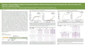 科学海报BrainPhys™ Neuronal Medium Supports the Electrical Activities of Neurons Derived from Human Pluripotent Stem Cells and Primary CNS Tissues in Long-Term Cultures


 沪公网安备31010102008431号