
 EasySep™小鼠TIL(CD45)正选试剂盒


技术资料
-
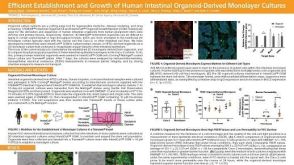 科学海报Efficient Establishment and Growth of Human Intestinal Organoid-Derived Monolayers
Conference:
ISSCR 2019
发布日期: 07/23/2019 -
-
-
 专家访谈Heather McCauley, PhD Improving Knowledge of Childhood Malabsorptive Disease with PSC-Derived Intestinal Organoids
研究方向:
上皮细胞生物学,类器官
发布日期: 07/06/2018 -
-
 专家访谈Luigi Aloia, PhD How Organoids are Driving Better Understanding of Liver Regeneration and Repair
研究方向:
上皮细胞生物学,类器官
发布日期: 04/11/2018


 沪公网安备31010102008431号