A triple co-culture model of the human respiratory tract to study immune-modulatory effects of liposomes and virosomes
The respiratory tract with its ease of access,vast surface area and dense network of antigen-presenting cells (APCs) represents an ideal target for immune-modulation. Bio-mimetic nanocarriers such as virosomes may provide immunomodulatory properties to treat diseases such as allergic asthma. In our study we employed a triple co-culture model of epithelial cells,macrophages and dendritic cells to simulate the human airway barrier. The epithelial cell line 16HBE was grown on inserts and supplemented with human blood monocyte-derived macrophages (MDMs) and dendritic cells (MDDCs) for exposure to influenza virosomes and liposomes. Additionally,primary human nasal epithelial cells (PHNEC) and EpCAM+ epithelial progenitor cell mono-cultures were utilized to simulate epithelium from large and smaller airways,respectively. To assess particle uptake and phenotype change,cell cultures were analyzed by flow cytometry and pro-inflammatory cytokine concentrations were measured by ELISA. All cell types internalized virosomes more efficiently than liposomes in both mono- and co-cultures. APCs like MDMs and MDDCs showed the highest uptake capacity. Virosome and liposome treatment caused a moderate degree of activation in MDDCs from mono-cultures and induced an increased cytokine production in co-cultures. In epithelial cells,virosome uptake was increased compared to liposomes in both mono- and co-cultures with EpCAM+ epithelial progenitor cells showing highest uptake capacity. In conclusion,all cell types successfully internalized both nanocarriers with virosomes being taken up by a higher proportion of cells and at a higher rate inducing limited activation of MDDCs. Thus virosomes may represent ideal carrier antigen systems to modulate mucosal immune responses in the respiratory tract without causing excessive inflammatory changes.
View Publication
产品号#:
05001
05021
05022
产品名:
PneumaCult™-ALI 培养基
PneumaCult™-ALI 培养基含12 mm Transwell®插件
PneumaCult™-ALI 培养基含6.5 mm Transwell®插件
Griggs TF et al. ( 2017)
Respiratory research 18 1 84
Rhinovirus C targets ciliated airway epithelial cells.
BACKGROUND The Rhinovirus C (RV-C),first identified in 2006,produce high symptom burdens in children and asthmatics,however,their primary target host cell in the airways remains unknown. Our primary hypotheses were that RV-C target ciliated airway epithelial cells (AECs),and that cell specificity is determined by restricted and high expression of the only known RV-C cell-entry factor,cadherin related family member 3 (CDHR3). METHODS RV-C15 (C15) infection in differentiated human bronchial epithelial cell (HBEC) cultures was assessed using immunofluorescent and time-lapse epifluorescent imaging. Morphology of C15-infected differentiated AECs was assessed by immunohistochemistry. RESULTS C15 produced a scattered pattern of infection,and infected cells were shed from the epithelium. The percentage of cells infected with C15 varied from 1.4 to 14.7% depending on cell culture conditions. Infected cells had increased staining for markers of ciliated cells (acetylated-alpha-tubulin [aat],p < 0.001) but not markers of goblet cells (wheat germ agglutinin or Muc5AC,p = ns). CDHR3 expression was increased on ciliated epithelial cells,but not other epithelial cells (p < 0.01). C15 infection caused a 27.4% reduction of ciliated cells expressing CDHR3 (p < 0.01). During differentiation of AECs,CDHR3 expression progressively increased and correlated with both RV-C binding and replication. CONCLUSIONS The RV-C only replicate in ciliated AECs in vitro,leading to infected cell shedding. CDHR3 expression positively correlates with RV-C binding and replication,and is largely confined to ciliated AECs. Our data imply that factors regulating differentiation and CDHR3 production may be important determinants of RV-C illness severity.
View Publication
产品号#:
05001
05021
05022
产品名:
PneumaCult™-ALI 培养基
PneumaCult™-ALI 培养基含12 mm Transwell®插件
PneumaCult™-ALI 培养基含6.5 mm Transwell®插件
Herawati E et al. ( 2016)
Journal of Cell Biology 214 5 571--586
Multiciliated cell basal bodies align in stereotypical patterns coordinated by the apical cytoskeleton
Multiciliated cells (MCCs) promote fluid flow through coordinated ciliary beating,which requires properly organized basal bodies (BBs). Airway MCCs have large numbers of BBs,which are uniformly oriented and,as we show here,align linearly. The mechanism for BB alignment is unexplored. To study this mechanism,we developed a long-term and high-resolution live-imaging system and used it to observe green fluorescent protein"centrin2"labeled BBs in cultured mouse tracheal MCCs. During MCC differentiation,the BB array adopted four stereotypical patterns,from a clustering floret? pattern to the linear alignment.? This alignment process was correlated with BB orientations,revealed by double immunostaining for BBs and their asymmetrically associated basal feet (BF). The BB alignment was disrupted by disturbing apical microtubules with nocodazole and by a BF-depleting Odf2 mutation. We constructed a theoretical model,which indicated that the apical cytoskeleton,acting like a viscoelastic fluid,provides a self-organizing mechanism in tracheal MCCs to align BBs linearly for mucociliary transport.
View Publication
Y. Bhattarai et al. (JUN 2018)
Cell host & microbe 23 6 775--785.e5
Gut Microbiota-Produced Tryptamine Activates an Epithelial G-Protein-Coupled Receptor to Increase Colonic Secretion.
Tryptamine,a tryptophan-derived monoamine similar to 5-hydroxytryptamine (5-HT),is produced by gut bacteria and is abundant in human and rodent feces. However,the physiologic effect of tryptamine in the gastrointestinal (GI) tract remains unknown. Here,we show that the biological effects of tryptamine are mediated through the 5-HT4 receptor (5-HT4R),a G-protein-coupled receptor (GPCR) uniquely expressed in the colonic epithelium. Tryptamine increases both ionic flux across the colonic epithelium and fluid secretion in colonoids from germ-free (GF) and humanized (ex-GF colonized with human stool) mice,consistent with increased intestinal secretion. The secretory effect of tryptamine is dependent on 5-HT4R activation and is blocked by 5-HT4R antagonist and absent in 5-HT4R-/- mice. GF mice colonized by Bacteroides thetaiotaomicron engineered to produce tryptamine exhibit accelerated GI transit. Our study demonstrates an aspect of host physiology under control of a bacterial metabolite that can be exploited as a therapeutic modality. VIDEO ABSTRACT.
View Publication
产品号#:
06005
产品名:
IntestiCult™ 类器官生长培养基 (小鼠)
M. D. Hu et al. (JUL 2018)
Journal of immunology (Baltimore,Md. : 1950) 201 2 747--756
Epithelial IL-15 Is a Critical Regulator of gamma$delta$ Intraepithelial Lymphocyte Motility within the Intestinal Mucosa.
Intraepithelial lymphocytes (IELs) expressing the gamma$delta$ TCR (gamma$delta$ IELs) provide continuous surveillance of the intestinal epithelium. However,the mechanisms regulating the basal motility of these cells within the epithelial compartment have not been well defined. We investigated whether IL-15 contributes to gamma$delta$ IEL localization and migratory behavior in addition to its role in IEL differentiation and survival. Using advanced live cell imaging techniques in mice,we find that compartmentalized overexpression of IL-15 in the lamina propria shifts the distribution of gamma$delta$ T cells from the epithelial compartment to the lamina propria. This mislocalization could be rescued by epithelial IL-15 overexpression,indicating that epithelial IL-15 is essential for gamma$delta$ IEL migration into the epithelium. Furthermore,in vitro analyses demonstrated that exogenous IL-15 stimulates gamma$delta$ IEL migration into cultured epithelial monolayers,and inhibition of IL-2Rbeta$ significantly attenuates the basal motility of these cells. Intravital microscopy showed that impaired IL-2Rbeta$ signaling induced gamma$delta$ IEL idling within the lateral intercellular space,which resulted in increased early pathogen invasion. Similarly,the redistribution of gamma$delta$ T cells to the lamina propria due to local IL-15 overproduction also enhanced bacterial translocation. These findings thus reveal a novel role for IL-15 in mediating gamma$delta$ T cell localization within the intestinal mucosa and regulating gamma$delta$ IEL motility and patrolling behavior as a critical component of host defense.
View Publication
产品号#:
06005
产品名:
IntestiCult™ 类器官生长培养基 (小鼠)
C. L. Kraft et al. (NOV 2017)
Oncotarget 8 61 102923--102933
GUCY2C maintains intestinal LGR5+stem cells by opposing ER stress.
Long-lived multipotent stem cells (ISCs) at the base of intestinal crypts adjust their phenotypes to accommodate normal maintenance and post-injury regeneration of the epithelium. Their long life,lineage plasticity,and proliferative potential underlie the necessity for tight homeostatic regulation of the ISC compartment. In that context,the guanylate cyclase C (GUCY2C) receptor and its paracrine ligands regulate intestinal epithelial homeostasis,including proliferation,lineage commitment,and DNA damage repair. However,a role for this axis in maintaining ISCs remains unknown. Transgenic mice enabling analysis of ISCs (Lgr5-GFP) in the context of GUCY2C elimination (Gucy2c -/- ) were combined with immunodetection techniques and pharmacological treatments to define the role of the GUCY2C signaling axis in supporting ISCs. ISCs were reduced inGucy2c -/- mice,associated with loss of active Lgr5+cells but a reciprocal increase in reserve Bmi1+cells. GUCY2C was expressed in crypt base Lgr5+cells in which it mediates canonical cyclic (c) GMP-dependent signaling. Endoplasmic reticulum (ER) stress,typically absent from ISCs,was elevated throughout the crypt base inGucy2c -/- mice. The chemical chaperone tauroursodeoxycholic acid resolved this ER stress and restored the balance of ISCs,an effect mimicked by the GUCY2C effector 8Br-cGMP. Reduced ISCs inGucy2c -/- mice was associated with greater epithelial injury and impaired regeneration following sub-lethal doses of irradiation. These observations suggest that GUCY2C provides homeostatic signals that modulate ER stress and cell vulnerability as part of the machinery contributing to the integrity of ISCs.
View Publication
产品号#:
06005
产品名:
IntestiCult™ 类器官生长培养基 (小鼠)
B. Wang et al. (FEB 2018)
Cell stem cell 22 2 206--220.e4
Phospholipid Remodeling and Cholesterol Availability Regulate Intestinal Stemness and Tumorigenesis.
Adequate availability of cellular building blocks,including lipids,is a prerequisite for cellular proliferation,but excess dietary lipids are linked to increased cancer risk. Despite these connections,specific regulatory relationships between membrane composition,intestinal stem cell (ISC) proliferation,and tumorigenesis are unclear. We reveal an unexpected link between membrane phospholipid remodeling and cholesterol biosynthesis and demonstrate that cholesterol itself acts as a mitogen for ISCs. Inhibition of the phospholipid-remodeling enzyme Lpcat3 increases membrane saturation and stimulates cholesterol biosynthesis,thereby driving ISC proliferation. Pharmacologic inhibition of cholesterol synthesis normalizes crypt hyperproliferation in Lpcat3-deficient organoids and mice. Conversely,increasing cellular cholesterol content stimulates crypt organoid growth,and providing excess dietary cholesterol or driving endogenous cholesterol synthesis through SREBP-2 expression promotes ISC proliferation in vivo. Finally,disruption of Lpcat3-dependent phospholipid and cholesterol homeostasis dramatically enhances tumor formation in Apcminmice. These findings identify a critical dietary-responsive phospholipid-cholesterol axis regulating ISC proliferation and tumorigenesis.
View Publication
NLRC3 is an inhibitory sensor of PI3K-mTOR pathways in cancer.
NLRs (nucleotide-binding domain and leucine-rich repeats) belong to a large family of cytoplasmic sensors that regulate an extraordinarily diverse range of biological functions. One of these functions is to contribute to immunity against infectious diseases,but dysregulation of their functional activity leads to the development of inflammatory and autoimmune diseases. Cytoplasmic innate immune sensors,including NLRs,are central regulators of intestinal homeostasis. NLRC3 (also known as CLR16.2 or NOD3) is a poorly characterized member of the NLR family and was identified in a genomic screen for genes encoding proteins bearing leucine-rich repeats (LRRs) and nucleotide-binding domains. Expression of NLRC3 is drastically reduced in the tumour tissue of patients with colorectal cancer compared to healthy tissues,highlighting an undefined potential function for this sensor in the development of cancer. Here we show that mice lacking NLRC3 are hyper-susceptible to colitis and colorectal tumorigenesis. The effect of NLRC3 is most dominant in enterocytes,in which it suppresses activation of the mTOR signalling pathways and inhibits cellular proliferation and stem-cell-derived organoid formation. NLRC3 associates with PI3Ks and blocks activation of the PI3K-dependent kinase AKT following binding of growth factor receptors or Toll-like receptor 4. These findings reveal a key role for NLRC3 as an inhibitor of the mTOR pathways,mediating protection against colorectal cancer.
View Publication
产品号#:
06005
产品名:
IntestiCult™ 类器官生长培养基 (小鼠)
Okkelman IA et al. ( 2016)
PloS one 11 12 e0167385
Use of Fluorescence Lifetime Imaging Microscopy (FLIM) as a Timer of Cell Cycle S Phase.
Incorporation of thymidine analogues in replicating DNA,coupled with antibody and fluorophore staining,allows analysis of cell proliferation,but is currently limited to monolayer cultures,fixed cells and end-point assays. We describe a simple microscopy imaging method for live real-time analysis of cell proliferation,S phase progression over several division cycles,effects of anti-proliferative drugs and other applications. It is based on the prominent (˜ 1.7-fold) quenching of fluorescence lifetime of a common cell-permeable nuclear stain,Hoechst 33342 upon the incorporation of 5-bromo-2'-deoxyuridine (BrdU) in genomic DNA and detection by fluorescence lifetime imaging microscopy (FLIM). We show that quantitative and accurate FLIM technique allows high-content,multi-parametric dynamic analyses,far superior to the intensity-based imaging. We demonstrate its uses with monolayer cell cultures,complex 3D tissue models of tumor cell spheroids and intestinal organoids,and in physiological study with metformin treatment.
View Publication

 EasySep™小鼠TIL(CD45)正选试剂盒


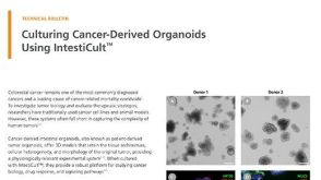
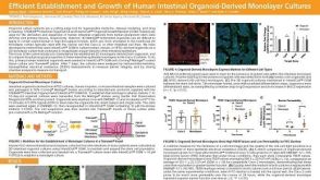 科学海报Efficient Establishment and Growth of Human Intestinal Organoid-Derived Monolayers

 沪公网安备31010102008431号