Jaatinen T et al. (MAR 2006)
Stem cells (Dayton,Ohio) 24 3 631--41
Global gene expression profile of human cord blood-derived CD133+ cells.
Human cord blood (CB)-derived CD133+ cells carry characteristics of primitive hematopoietic cells and proffer an alternative for CD34+ cells in hematopoietic stem cell (HSC) transplantation. To characterize the CD133+ cell population on a genetic level,a global expression analysis of CD133+ cells was performed using oligonucleotide microarrays. CD133+ cells were purified from four fresh CB units by immunomagnetic selection. All four CD133+ samples showed significant similarity in their gene expression pattern,whereas they differed clearly from the CD133- control samples. In all,690 transcripts were differentially expressed between CD133+ and CD133- cells. Of these,393 were increased and 297 were decreased in CD133+ cells. The highest overexpression was noted in genes associated with metabolism,cellular physiological processes,cell communication,and development. A set of 257 transcripts expressed solely in the CD133+ cell population was identified. Colony-forming unit (CFU) assay was used to detect the clonal progeny of precursors present in the studied cell populations. The results demonstrate that CD133+ cells express primitive markers and possess clonogenic progenitor capacity. This study provides a gene expression profile for human CD133+ cells. It presents a set of genes that may be used to unravel the properties of the CD133+ cell population,assumed to be highly enriched in HSCs.
View Publication
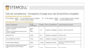
产品号#:
04434
04444
产品名:
MethoCult™ H4434 Classic
MethoCult™ H4434 Classic
Giassi LJ et al. (AUG 2008)
Experimental biology and medicine (Maywood,N.J.) 233 8 997--1012
Expanded CD34+ human umbilical cord blood cells generate multiple lymphohematopoietic lineages in NOD-scid IL2rgamma(null) mice.
Umbilical cord blood (UCB) is increasingly being used for human hematopoietic stem cell (HSC) transplantation in children but often requires pooling multiple cords to obtain sufficient numbers for transplantation in adults. To overcome this limitation,we have used an ex vivo two-week culture system to expand the number of hematopoietic CD34(+) cells in cord blood. To assess the in vivo function of these expanded CD34(+) cells,cultured human UCB containing 1 x 10(6) CD34(+) cells were transplanted into conditioned NOD-scid IL2rgamma(null) mice. The expanded CD34(+) cells displayed short- and long-term repopulating cell activity. The cultured human cells differentiated into myeloid,B-lymphoid,and erythroid lineages,but not T lymphocytes. Administration of human recombinant TNFalpha to recipient mice immediately prior to transplantation promoted human thymocyte and T-cell development. These T cells proliferated vigorously in response to TCR cross-linking by anti-CD3 antibody. Engrafted TNFalpha-treated mice generated antibodies in response to T-dependent and T-independent immunization,which was enhanced when mice were co-treated with the B cell cytokine BLyS. Ex vivo expanded CD34(+) human UCB cells have the capacity to generate multiple hematopoietic lineages and a functional human immune system upon transplantation into TNFalpha-treated NOD-scid IL2rgamma(null) mice.
View Publication
Angiopoietin-like 5 and IGFBP2 stimulate ex vivo expansion of human cord blood hematopoietic stem cells as assayed by NOD/SCID transplantation.
Hematopoietic stem cells (HSCs) are the basis of bone marrow transplantation and are attractive target cells for hematopoietic gene therapy,but these important clinical applications have been severely hampered by difficulties in ex vivo expansion of HSCs. In particular,the use of cord blood for adult transplantation is greatly limited by the number of HSCs. Previously we identified angiopoietin-like proteins and IGF-binding protein 2 (IGFBP2) as new hormones that,together with other factors,can expand mouse bone marrow HSCs in culture. Here,we measure the activity of multipotent human severe combined immunodeficient (SCID)-repopulating cells (SRCs) by transplantation into the nonobese diabetic SCID (NOD/SCID) mice; secondary transplantation was performed to evaluate the self-renewal potential of SRCs. A serum-free medium containing SCF,TPO,and FGF-1 or Flt3-L cannot significantly support expansion of the SRCs present in human cord blood CD133+ cells. Addition of either angiopoietin-like 5 or IGF-binding protein 2 to the cultures led to a sizable expansion of HSC numbers,as assayed by NOD/SCID transplantation. A serum-free culture containing SCF,TPO,FGF-1,angiopoietin-like 5,and IGFBP2 supports an approximately 20-fold net expansion of repopulating human cord blood HSCs,a number potentially applicable to several clinical processes including HSC transplantation.
View Publication
产品号#:
09600
09650
28600
产品名:
StemSpan™ SFEM
StemSpan™ SFEM
L-Calc™有限稀释软件
Lin H et al. (MAR 2009)
Experimental biology and medicine (Maywood,N.J.) 234 3 342--53
Maitake beta-glucan enhances umbilical cord blood stem cell transplantation in the NOD/SCID mouse.
Beta glucans are cell wall constituents of yeast,fungi and bacteria,as well as mushrooms and barley. Glucans are not expressed on mammalian cells and are recognized as pathogen-associated molecular patterns (PAMPS) by pattern recognition receptors (PRR). Beta glucans have potential activity as biological response modifiers for hematopoiesis and enhancement of bone marrow recovery after injury. We have reported that Maitake beta glucan (MBG) enhanced mouse bone marrow (BMC) and human umbilical cord blood (CB) cell granulocyte-monocyte colony forming unit (GM-CFU) activity in vitro and protected GM-CFU forming stem cells from doxorubicin (DOX) toxicity. The objective of this study was to determine the effects of MBG on expansion of phenotypically distinct subpopulations of progenitor and stem cells in CB from full-term infants cultured ex vivo and on homing and engraftment in vivo in the nonobese diabetic/severe combined immunodeficient (NOD/SCID) mouse. MBG promoted a greater expansion of CD34+CD33+CD38- human committed hematopoietic progenitor (HPC) cells compared to the conventional stem cell culture medium (P = 0.002 by ANOVA). CD34+CXCR4+CD38- early,uncommitted human hematopoietic stem cell (HSC) numbers showed a trend towards increase in response to MBG. The fate of CD34+ enriched CB cells after injection into the sublethally irradiated NOS/SCID mouse was evaluated after retrieval of xenografted human CB from marrow and spleen by flow cytometric analysis. Oral administration of MBG to recipient NOS/SCID mice led to enhanced homing at 3 days and engraftment at 6 days in mouse bone marrow (P = 0.002 and P = 0.0005,respectively) compared to control mice. More CD34+ human CB cells were also retrieved from mouse spleen in MBG treated mice at 6 days after transplantation. The studies suggest that MBG promotes hematopoiesis through effects on CD34+ progenitor cell expansion ex vivo and when given to the transplant recipient could enhance CD34+ precursor cell homing and support engraftment.
View Publication
产品号#:
02690
09600
09650
09850
15026
15066
产品名:
StemSpan™ CC100
StemSpan™ SFEM
StemSpan™ SFEM
RosetteSep™人造血祖细胞富集抗体混合物
RosetteSep™人造血祖细胞富集抗体混合物
Xia L et al. (NOV 2004)
Blood 104 10 3091--6
Surface fucosylation of human cord blood cells augments binding to P-selectin and E-selectin and enhances engraftment in bone marrow.
Murine hematopoietic stem and progenitor cells (HSPCs) home to bone marrow in part by rolling on P-selectin and E-selectin expressed on endothelial cells. Human adult CD34(+) cells,which are enriched in HSPCs,roll on endothelial selectins in bone marrow vessels of nonobese diabetic/severe combined immune deficiency (NOD/SCID) mice. Many human umbilical cord blood (CB) CD34(+) cells do not roll in these vessels,in part because of an uncharacterized defect in binding to P-selectin. Selectin ligands must be alpha1-3 fucosylated to form glycan determinants such as sialyl Lewis x (sLe(x)). We found that inadequate alpha1-3 fucosylation of CB CD34(+) cells,particularly CD34(+)CD38(-/low) cells that are highly enriched in HSPCs,caused them to bind poorly to E-selectin as well as to P-selectin. Treatment of CB CD34(+) cells with guanosine diphosphate (GDP) fucose and exogenous alpha1-3 fucosyltransferase VI increased cell-surface sLe(x) determinants,augmented binding to fluid-phase P- and E-selectin,and improved cell rolling on P- and E-selectin under flow. Similar treatment of CB mononuclear cells enhanced engraftment of human hematopoietic cells in bone marrows of irradiated NOD/SCID mice. These observations suggest that alpha1-3 fucosylation of CB cells might be a simple and effective method to improve hematopoietic cell homing to and engraftment in bone marrows of patients receiving CB transplants.
View Publication
产品号#:
产品名:
Flores-Figueroa E et al. (FEB 2005)
Leukemia research 29 2 215--24
Mesenchymal stem cells in myelodysplastic syndromes: phenotypic and cytogenetic characterization.
Bone marrow-derived mesenchymal stem cells (MSC) have been defined as primitive,undifferentiated cells,capable of self-renewal and with the ability to give rise to different cell lineages,including adipocytes,osteocytes,fibroblasts,chondrocytes,and myoblasts. MSC are key components of the hematopoietic microenvironment. Several studies,including some from our own group,suggest that important quantitative and functional alterations are present in the stroma of patients with myelodysplasia (MDS). However,in most of such studies the stroma has been analyzed as a complex network of different cell types and molecules,thus it has been difficult to identify and characterize the cell(s) type(s) that is (are) altered in MDS. In the present study,we have focused on the biological characterization of MSC from MDS. As a first approach,we have quantified their numbers in bone marrow,and have worked on their phenotypic (morphology and immunophenotype) and cytogenetic properties. MSC were obtained by a negative selection procedure and cultured in a MSC liquid culture medium. In terms of morphology,as well as the expression of certain cell markers,no differences were observed between MSC from MDS patients and those derived from normal marrow. In both cases,MSC expressed CD29,CD90,CD105 and Prolyl-4-hydroxylase; in contrast,they did not express CD14,CD34,CD68,or alkaline phosphatase. Interestingly,in five out of nine MDS patients,MSC developed in culture showed cytogenetic abnormalities,usually involving the loss of chromosomal material. All those five cases also showed cytogenetic abnormalities in their hematopoietic cells. Interestingly,in some cases there was a complete lack of overlap between the karyotypes of hematopoietic cells and MSC. To the best of our knowledge,the present study is the first in which a pure population of MSC from MDS patients is analyzed in terms of their whole karyotype and demonstrates that in a significant proportion of patients,MSC are cytogenetically abnormal. Although the reason of this is still unclear,such alterations may have an impact on the physiology of these cells. Further studies are needed to assess the functional integrity of MDS-derived MSC.
View Publication

 EasySep™小鼠TIL(CD45)正选试剂盒


 电子书血液样本制备
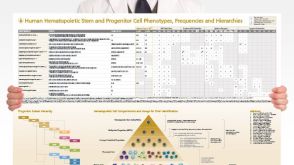 挂图Human Hematopoietic Stem and Progenitor Cell Phenotyping Overview of subset surface markers, frequencies and assays for analysis

 沪公网安备31010102008431号