Lam AC et al. (DEC 2001)
Transfusion 41 12 1567--76
Preclinical ex vivo expansion of cord blood hematopoietic stem and progenitor cells: duration of culture; the media, serum supplements, and growth factors used; and engraftment in NOD/SCID mice.
BACKGROUND: Ex vivo expansion of cord blood (CB) hematopoietic stem and progenitor cells increases cell dose and may reduce the severity and duration of neutropenia and thrombocytopenia after transplantation. This study's purpose was to establish a clinically applicable culture system by investigating the use of cytokines,serum-free media,and autologous plasma for the expansion of CB cells and the engraftment of expanded product in nonobese diabetic/severe combined immunodeficient (NOD/SCID) mice. STUDY DESIGN AND METHODS: Enriched CB CD34+ cells were cultured in four media (Iscove's modified Dulbecco's medium with FCS,Gibco; X-Vivo-10,BioWhittaker; QBSF-60,Quality Biological; and StemSpan SFEM,Stem Cell Technologies) with four cytokine combinations (thrombopoietin [TPO],SCF,Flt-3 ligand [FL] with and without G-CSF,and/or IL-6). The effect of autologous CB plasma was also investigated. The read-out measures were evaluated on Days 8 and 12. After expansion at the optimized condition,cultured cells were transplanted into sublethally irradiated NOD/SCID mice. The engraftment of human CD45+ cells and subsets in the bone marrow,spleen,and peripheral blood was determined. RESULTS: QBSF-60 or StemSpan SFEM supported high yields of early progenitors (CD34+ cells,textlessor= 64.8-fold; CD34+CD38- cells,330-fold; CFU-granulocyte erythroid macrophage megakaryocyte [GEMM],248-fold) and CFUs of the myeloid (CFU-GM,407-fold) and erythroid (BFU/CFU-E,144-fold) lineages. The expansion of the megakaryocytic lineage was consistently higher in X-Vivo-10 (CFU-megakaryocyte,684-fold). Autologous plasma promoted colony formation but reduced CD34+ cells and CFU-GEMM. The addition of G-CSF or IL-6 improved cell yields; G-CSF was more effective for committed progenitors. Expansion products from cultures in QBSF-60 with the cytokines engrafted and differentiated into the myeloid and lymphoid lineages in NOD/SCID mice. CONCLUSION: The data supported the strategy of expansion. The optimized condition may be applicable to clinical expansion for the abrogation or reduction of posttransplant cytopenia.
View Publication
产品号#:
09600
09650
产品名:
StemSpan™ SFEM
StemSpan™ SFEM
Schiedlmeier B et al. (MAR 2003)
Blood 101 5 1759--68
High-level ectopic HOXB4 expression confers a profound in vivo competitive growth advantage on human cord blood CD34+ cells, but impairs lymphomyeloid differentiation.
Ectopic retroviral expression of homeobox B4 (HOXB4) causes an accelerated and enhanced regeneration of murine hematopoietic stem cells (HSCs) and is not known to compromise any program of lineage differentiation. However,HOXB4 expression levels for expansion of human stem cells have still to be established. To test the proposed hypothesis that HOXB4 could become a prime tool for in vivo expansion of genetically modified human HSCs,we retrovirally overexpressed HOXB4 in purified cord blood (CB) CD34+ cells together with green fluorescent protein (GFP) as a reporter protein,and evaluated the impact of ectopic HOXB4 expression on proliferation and differentiation in vitro and in vivo. When injected separately into nonobese diabetic-severe combined immunodeficient (NOD/SCID) mice or in competition with control vector-transduced cells,HOXB4-overexpressing cord blood CD34+ cells had a selective growth advantage in vivo,which resulted in a marked enhancement of the primitive CD34+ subpopulation (P =.01). However,high HOXB4 expression substantially impaired the myeloerythroid differentiation program,and this was reflected in a severe reduction of erythroid and myeloid progenitors in vitro (P textless.03) and in vivo (P =.01). Furthermore,HOXB4 overexpression also significantly reduced B-cell output (P textless.01). These results show for the first time unwanted side effects of ectopic HOXB4 expression and therefore underscore the need to carefully determine the therapeutic window of HOXB4 expression levels before initializing clinical trials.
View Publication
产品号#:
04434
04444
09600
09650
产品名:
MethoCult™ H4434 Classic
MethoCult™ H4434 Classic
StemSpan™ SFEM
StemSpan™ SFEM
Rank G et al. (SEP 2010)
Blood 116 9 1585--92
Identification of a PRMT5-dependent repressor complex linked to silencing of human fetal globin gene expression.
Defining the molecular mechanisms underpinning fetal (gamma) globin gene silencing may provide strategies for reactivation of gamma-gene expression,a major therapeutic objective in patients with beta-thalassemia and sickle cell disease (SCD). We have previously demonstrated that symmetric methylation of histone H4 Arginine 3 (H4R3me2s) by the protein arginine methyltransferase PRMT5 is required for recruitment of the DNA methyltransferase DNMT3A to the gamma-promoter,and subsequent DNA methylation and gene silencing. Here we show in an erythroid cell line,and in primary adult erythroid progenitors that PRMT5 induces additional repressive epigenetic marks at the gamma-promoter through the assembly of a multiprotein repressor complex containing the histone modifying enzymes SUV4-20h1,casein kinase 2alpha (CK2alpha),and components of the nucleosome remodeling and histone deacetylation complex. Expression of a mutant form of PRMT5 lacking methyltransferase activity or shRNA-mediated knockdown of SUV4-20h1 resulted in loss of complex binding to the gamma-promoter,reversal of both histone and DNA repressive epigenetic marks,and increased gamma-gene expression. The repressive H4K20me3 mark induced by SUV4-20h1 is enriched on the gamma-promoter in erythroid progenitors from adult bone marrow compared with cord blood,suggesting developmental specificity. These studies define coordinated epigenetic events linked to fetal globin gene silencing,and provide potential therapeutic targets for the treatment of beta-thalassemia and SCD.
View Publication
产品号#:
09600
09650
产品名:
StemSpan™ SFEM
StemSpan™ SFEM
Ito CY et al. (JAN 2010)
Blood 115 2 257--60
The AC133+CD38-, but not the rhodamine-low, phenotype tracks LTC-IC and SRC function in human cord blood ex vivo expansion cultures.
Phenotypic markers associated with human hematopoietic stem cells (HSCs) were developed and validated using uncultured cells. Because phenotype and function can be dissociated during culture,better markers to prospectively track and isolate HSCs in ex vivo cultures could be instrumental in advancing HSC-based therapies. Using an expansion system previously shown to increase hematopoietic progenitors and SCID-repopulating cells (SRCs),we demonstrated that the rhodamine-low phenotype was lost,whereas AC133 expression was retained throughout culture. Furthermore,the AC133(+)CD38(-) subpopulation was significantly enriched in long-term culture-initiating cells (LTC-IC) and SRCs after culture. Preculture and postculture analysis of total nucleated cell and LTC-IC number,and limiting dilution analysis in NOD/SCID mice,showed a 43-fold expansion of the AC133(+)CD38(-) subpopulation that corresponded to a 7.3-fold and 4.4-fold expansion of LTC-ICs and SRCs in this subpopulation,respectively. Thus,AC133(+)CD38(-) is an improved marker that tracks and enriches for LTC-IC and SRC in ex vivo cultures.
View Publication
Pesce M et al. (SEP 2003)
Circulation research 93 5 e51--62
Myoendothelial differentiation of human umbilical cord blood-derived stem cells in ischemic limb tissues.
Human umbilical cord blood (UCB) contains high numbers of endothelial progenitors cells (EPCs) characterized by coexpression of CD34 and CD133 markers. Prior studies have shown that CD34+/CD133+ EPCs from the cord or peripheral blood (PB) can give rise to endothelial cells and induce angiogenesis in ischemic tissues. In the present study,it is shown that freshly isolated human cord blood CD34+ cells injected into ischemic adductor muscles gave rise to endothelial and,unexpectedly,to skeletal muscle cells in mice. In fact,the treated limbs exhibited enhanced arteriole length density and regenerating muscle fiber density. Under similar experimental conditions,CD34- cells did not enhance the formation of new arterioles and regenerating muscle fibers. In nonischemic limbs CD34+ cells increased arteriole length density but did not promote formation of new muscle fibers. Endothelial and myogenic differentiation ability was maintained in CD34+ cells after ex vivo expansion. Myogenic conversion of human cord blood CD34+ cells was also observed in vitro by coculture onto mouse myoblasts. These results show that human cord blood CD34+ cells differentiate into endothelial and skeletal muscle cells,thus providing an indication of human EPCs plasticity. The full text of this article is available online at http://www.circresaha.org.
View Publication
产品号#:
09600
09650
84535
84545
产品名:
StemSpan™ SFEM
StemSpan™ SFEM
Ioannidis P et al. (MAY 2005)
The Journal of biological chemistry 280 20 20086--93
CRD-BP/IMP1 expression characterizes cord blood CD34+ stem cells and affects c-myc and IGF-II expression in MCF-7 cancer cells.
The coding region determinant-binding protein/insulin-like growth factor II mRNA-binding protein (CRD-BP/IMP1) is an RNA-binding protein specifically recognizing c-myc,leader 3' IGF-II and tau mRNAs,and the H19 RNA. CRD-BP/IMP1 is predominantly expressed in embryonal tissues but is de novo activated and/or overexpressed in various human neoplasias. To address the question of whether CRD-BP/IMP1 expression characterizes certain cell types displaying distinct proliferation and/or differentiation properties (i.e. stem cells),we isolated cell subpopulations from human bone marrow,mobilized peripheral blood,and cord blood,all sources known to contain stem cells,and monitored for its expression. CRD-BP/IMP1 was detected only in cord blood-derived CD34(+) stem cells and not in any other cell type of either adult or cord blood origin. Adult BM CD34(+) cells cultured in the presence of 5'-azacytidine expressed de novo CRD-BP/IMP1,suggesting that epigenetic modifications may be responsible for its silencing in adult non-expressing cells. Furthermore,by applying the short interfering RNA methodology in MCF-7 cells,we observed,subsequent to knocking down CRD-BP/IMP1,decreased c-myc expression,increased IGF-II mRNA levels,and reduced cell proliferation rates. These data 1) suggest a normal role for CRD-BP/IMP1 in pluripotent stem cells with high renewal capacity,like the CB CD34(+) cells,2) indicate that altered methylation may directly or indirectly affect its expression in adult cells,3) imply that its de novo activation in cancer cells may affect the expression of c-Myc and insulin-like growth factor II,and 4) indicate that the inhibition of CRD-BP/IMP1 expression might affect cancer cell proliferation.
View Publication

 EasySep™小鼠TIL(CD45)正选试剂盒


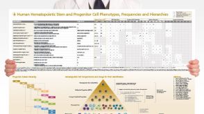 挂图Human Hematopoietic Stem and Progenitor Cell Phenotyping Overview of subset surface markers, frequencies and assays for analysis


 沪公网安备31010102008431号