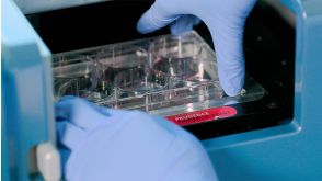
Reconstitution of the functional human hematopoietic microenvironment derived from human mesenchymal stem cells in the murine bone marrow compartment.
Hematopoiesis is maintained by specific interactions between both hematopoietic and nonhematopoietic cells. Whereas hematopoietic stem cells (HSCs) have been extensively studied both in vitro and in vivo,little is known about the in vivo characteristics of stem cells of the nonhematopoietic component,known as mesenchymal stem cells (MSCs). Here we have visualized and characterized human MSCs in vivo following intramedullary transplantation of enhanced green fluorescent protein-marked human MSCs (eGFP-MSCs) into the bone marrow (BM) of nonobese diabetic/severe combined immunodeficiency (NOD/SCID) mice. Between 4 to 10 weeks after transplantation,eGFP-MSCs that engrafted in murine BM integrated into the hematopoietic microenvironment (HME) of the host mouse. They differentiated into pericytes,myofibroblasts,BM stromal cells,osteocytes in bone,bone-lining osteoblasts,and endothelial cells,which constituted the functional components of the BM HME. The presence of human MSCs in murine BM resulted in an increase in functionally and phenotypically primitive human hematopoietic cells. Human MSC-derived cells that reconstituted the HME appeared to contribute to the maintenance of human hematopoiesis by actively interacting with primitive human hematopoietic cells.
View Publication
产品号#:
04034
04044
产品名:
MethoCult™ H4034 Optimum
MethoCult™ H4034 Optimum
Kang YK et al. (MAR 2016)
Blood research 51 1 31--6
Humanizing NOD/SCID/IL-2Rγnull (NSG) mice using busulfan and retro-orbital injection of umbilical cord blood-derived CD34(+) cells.
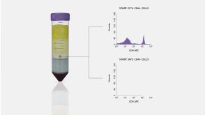
BACKGROUND Humanized mouse models are still under development,and various protocols exist to improve human cell engraftment and function. METHODS Fourteen NOD/SCID/IL-2Rγnull (NSG) mice (4‒5 wk old) were conditioned with busulfan and injected with human umbilical cord blood (hUCB)-derived CD34(+) hematopoietic stem cells (HSC) via retro-orbital sinuses. The bone marrow (BM),spleen,and peripheral blood (PB) were analyzed 8 and 12 weeks after HSC transplantation. RESULTS Most of the NSG mice tolerated the regimen well. The percentage of hCD45(+) and CD19(+) cells rose significantly in a time-dependent manner. The median percentage of hCD45(+)cells in the BM was 55.5% at week 8,and 67.2% at week 12. The median percentage of hCD45(+) cells in the spleen at weeks 8 and 12 was 42% and 51%,respectively. The median percentage of hCD19(+) cells in BM at weeks 8 and 12 was 21.5% and 39%,respectively (P=0.04). Similarly,the median percentage of hCD19(+) cells in the spleen at weeks 8 and 12 was 10% and 24%,respectively (P=0.04). The percentage of hCD19(+) B cells in PB was 23% at week 12. At week 8,hCD3(+) T cells were barely detectable,while hCD7(+) was detected in the BM and spleen. The percentage of hCD3(+) T cells was 2‒3% at week 12 in the BM,spleen,and PB of humanized NSG mice. CONCLUSION We adopted a simplified protocol for establishing humanized NSG mice. We observed a higher engraftment rate of human CD45(+) cells than earlier studies without any significant toxicity. And human CD45(+) cell engraftment at week 8 was comparable to that of week 12.
View Publication

 EasySep™小鼠TIL(CD45)正选试剂盒


 沪公网安备31010102008431号