Carlo-Stella C et al. (JAN 2007)
Stem cells (Dayton,Ohio) 25 1 252--61
Placental growth factor-1 potentiates hematopoietic progenitor cell mobilization induced by granulocyte colony-stimulating factor in mice and nonhuman primates.
The complex hematopoietic effects of placental growth factor (PlGF) prompted us to test in mice and nonhuman primates the mobilization of peripheral blood progenitor cells (PBPCs) elicited by recombinant mouse PlGF-2 (rmPlGF-2) and recombinant human PlGF-1 (rhPlGF-1). PBPC mobilization was evaluated by assaying colony-forming cells (CFCs),high-proliferative potential-CFCs (HPP-CFCs),and long-term culture-initiating cells (LTC-ICs). In mice,both rmPlGF-2 and rhPlGF-1 used as single agents failed to mobilize PBPCs,whereas the combination of rhPlGF-1 and granulocyte colony-stimulating factor (rhG-CSF) increased CFCs and LTC-ICs per milliliter of blood by four- and eightfold,respectively,as compared with rhG-CSF alone. rhPlGF-1 plus rhG-CSF significantly increased matrix metalloproteinase-9 plasma levels over rhG-CSF alone,suggesting a mechanistic explanation for rhPlGF-1/rhG-CSF synergism. In rhesus monkeys,rhPlGF-1 alone had no mobilization effect,whereas rhPlGF-1 (260 microg/kg per day) plus rhG-CSF (100 microg/kg per day) increased rhG-CSF-elicited mobilization of CFCs,HPP-CFCs,and LTC-ICs per milliliter of blood by 5-,7-,and 15-fold,respectively. No specific toxicity was associated with the administration of rhPlGF-1 alone or in combination. In conclusion,our data demonstrate that rhPlGF-1 significantly increases rhG-CSF-elicited hematopoietic mobilization and provide a preclinical rationale for evaluating rhPlGF-1 in the clinical setting.
View Publication
产品号#:
05150
产品名:
MyeloCult™ H5100
Wang Y et al. (MAR 2007)
Blood 109 5 2147--55
Adaptive secretion of granulocyte-macrophage colony-stimulating factor (GM-CSF) mediates imatinib and nilotinib resistance in BCR/ABL+ progenitors via JAK-2/STAT-5 pathway activation.
Overcoming imatinib mesylate (IM) resistance and disease persistence in patients with chronic myeloid leukemia (CML) is of considerable importance to the issue of potential cure. Here we asked whether autocrine signaling contributes to survival of BCR/ABL+ cells in the presence of IM and nilotinib (NI; AMN107),a novel,more selective Abl inhibitor. Conditioned media (CM) of IM-resistant LAMA84 cell clones (R-CM) was found to substantially protect IM-naive LAMA cells and primary CML progenitors from IM- or NI-induced cell death. This was due to an increased secretion of the granulocyte-macrophage colony-stimulating factor (GM-CSF),which was identified as the causative factor mediating IM resistance in R-CM. GM-CSF elicited IM and NI drug resistance via a BCR/ABL-independent activation of the janus kinases 2 (JAK-2)/signal transducer and activator of transcription 5 (STAT-5) signaling pathway in GM-CSF receptor alpha receptor (CD116)-expressing cells,including primary CD34+/CD116+ GM progenitors (GMPs). Elevated mRNA and protein levels of GM-CSF were detected in IM-resistant patient samples,suggesting a contribution of GM-CSF secretion for IM and NI resistance in vivo. Importantly,inhibition of JAK-2 with AG490 abrogated GM-CSF-mediated STAT-5 phosphorylation and NI resistance in vitro. Together,adaptive autocrine secretion of GM-CSF mediates BCR/ABL-independent IM and NI resistance via activation of the antiapoptotic JAK-2/STAT-5 pathway. Inhibition of JAK-2 overcomes GM-CSF-induced IM and NI progenitor cell resistance,providing a rationale for the application of JAK-2 inhibitors to eradicate residual disease in CML.
View Publication
产品号#:
04230
产品名:
MethoCult™ H4230
Feng R et al. (MAR 2007)
Blood 109 5 2130--8
SDX-308, a nonsteroidal anti-inflammatory agent, inhibits NF-kappaB activity, resulting in strong inhibition of osteoclast formation/activity and multiple myeloma cell growth.
Multiple myeloma is characterized by increased osteoclast activity that results in bone destruction and lytic lesions. With the prolonged overall patient survival achieved by new treatment modalities,additional drugs are required to inhibit bone destruction. We focused on a novel and more potent structural analog of the nonsteroidal anti-inflammatory drug etodolac,known as SDX-308,and its effects on osteoclastogenesis and multiple myeloma cells. SDX-101 is another structural analog of etodolac that is already used in clinical trials for the treatment of B-cell chronic lymphocytic leukemia (B-CLL). Compared with SDX-101,a 10-fold lower concentration of SDX-308 induced potent (60%-80%) inhibition of osteoclast formation,and a 10- to 100-fold lower concentration inhibited multiple myeloma cell proliferation. Bone resorption was completely inhibited by SDX-308,as determined in dentin-based bone resorption assays. SDX-308 decreased constitutive and RANKL-stimulated NF-kappaB activation and osteoclast formation in an osteoclast cellular model,RAW 264.7. SDX-308 effectively suppressed TNF-alpha-induced IKK-gamma and IkappaB-alpha phosphorylation and degradation and subsequent NF-kappaB activation in human multiple myeloma cells. These results indicate that SDX-308 effectively inhibits multiple myeloma cell proliferation and osteoclast activity,potentially by controlling NF-kappaB activation signaling. We propose that SDX-308 is a promising therapeutic candidate to inhibit multiple myeloma growth and osteoclast activity and that it should receive attention for further study.
View Publication
产品号#:
04434
04444
产品名:
MethoCult™ H4434 Classic
MethoCult™ H4434 Classic
Kubota Y et al. (MAR 2007)
Journal of immunology (Baltimore,Md. : 1950) 178 5 2923--31
Mcl-1 depletion in apoptosis elicited by ionizing radiation in peritoneal resident macrophages of C3H mice.
Remarkably,apoptosis was induced by exposing peritoneal resident macrophages (PRM) of C3H mice,but not other strains of mice,to ionizing radiation. The molecular mechanism of this strain-specific apoptosis in PRM was studied. The apoptosis elicited in C3H mouse PRM 4 h after exposure was effectively blocked by proteasome inhibitors. Irradiation-induced disruption of mitochondrial transmembrane potential and the release of cytochrome c into the cytosol were also suppressed by a proteasome inhibitor but not by a caspase inhibitor. To determine whether the apoptosis occurred due to a depletion of antiapoptotic proteins,Bcl-2 family proteins were examined. Irradiation markedly decreased the level of Mcl-1,but not Bcl-2,Bcl-X(L),Bax,A1,or cIAP1. Mcl-1's depletion was suppressed by a proteasome inhibitor but not by a caspase inhibitor. The amount of Mcl-1 was well correlated with the rate of apoptosis in C3H mouse PRM exposed to irradiation and not affected by irradiation in radioresistant B6 mouse PRM. Irradiation increased rather than decreased the Mcl-1 mRNA expression in C3H mouse PRM. On the other hand,Mcl-1 protein synthesis was markedly suppressed by irradiation. Global protein synthesis was also suppressed by irradiation in C3H mouse PRM but not in B6 mouse PRM. The down-regulation of Mcl-1 expression with Mcl-1-specific small interfering RNA or antisense oligonucleotide significantly induced apoptosis in both C3H and B6 mouse PRM without irradiation. It was concluded that the apoptosis elicited in C3H mouse PRM by ionizing radiation was attributable to the depletion of Mcl-1 through radiation-induced arrest of global protein synthesis.
View Publication
产品号#:
03534
产品名:
MethoCult™ GF M3534
Sjogren A-KM et al. (MAY 2007)
The Journal of clinical investigation 117 5 1294--304
GGTase-I deficiency reduces tumor formation and improves survival in mice with K-RAS-induced lung cancer.
Protein geranylgeranyltransferase type I (GGTase-I) is responsible for the posttranslational lipidation of CAAX proteins such as RHOA,RAC1,and cell division cycle 42 (CDC42). Inhibition of GGTase-I has been suggested as a strategy to treat cancer and a host of other diseases. Although several GGTase-I inhibitors (GGTIs) have been synthesized,they have very different properties,and the effects of GGTIs and GGTase-I deficiency are unclear. One concern is that inhibiting GGTase-I might lead to severe toxicity. In this study,we determined the effects of GGTase-I deficiency on cell viability and K-RAS-induced cancer development in mice. Inactivating the gene for the critical beta subunit of GGTase-I eliminated GGTase-I activity,disrupted the actin cytoskeleton,reduced cell migration,and blocked the proliferation of fibroblasts expressing oncogenic K-RAS. Moreover,the absence of GGTase-I activity reduced lung tumor formation,eliminated myeloproliferative phenotypes,and increased survival of mice in which expression of oncogenic K-RAS was switched on in lung cells and myeloid cells. Interestingly,several cell types remained viable in the absence of GGTase-I,and myelopoiesis appeared to function normally. These findings suggest that inhibiting GGTase-I may be a useful strategy to treat K-RAS-induced malignancies.
View Publication
Pessina A et al. (FEB 2009)
Toxicology in vitro : an international journal published in association with BIBRA 23 1 194--200
Application of human CFU-Mk assay to predict potential thrombocytotoxicity of drugs.
Megakaryocytopoiesis gives rise to platelets by proliferation and differentiation of lineage-specific progenitors,identified in vitro as Colony Forming Unit-Megakaryocytes (CFU-Mk). The aim of this study was to refine and optimize the in vitro Standard Operating Procedure (SOP) of the CFU-Mk assay for detecting drug-induced thrombocytopenia and to prevalidate a model for predicting the acute exposure levels that cause maximum tolerated decreases in the platelets count,based on the correlation with the maximal plasma concentrations (C max) in vivo. The assay was linear under the SOP conditions,and the in vitro endpoints (percentage of colonies growing) were reproducible within and across laboratories. The protocol performance phase was carried out testing 10 drugs (selected on the base of their recognised or potential in vivo haematotoxicity,according to the literature). Results showed that a relationship can be established between the maximal concentration in plasma (C max) and the in vitro concentrations that inhibited the 10-50-90 percent of colonies growth (ICs). When C max is lower than IC10,it is possible to predict that the chemicals have no direct toxicity effect on CFU-Mk and could not induce thrombocytopenia due to bone marrow damage. When the C max is higher than IC90 and/or IC50,thrombocytopenia can occur due to direct toxicity of chemicals on CFU-Mk progenitors.
View Publication
产品号#:
04960
04902
04900
04963
04962
04970
产品名:
MegaCult™-C胶原和无细胞因子培养基
胶原蛋白溶液
MegaCult™-C无细胞因子培养基
双室载玻片套件
MegaCult™-C CFU-Mk染色试剂盒
MegaCult™-C无细胞因子全套试剂盒
Karp JE et al. (MAY 2009)
Blood 113 20 4841--52
Active oral regimen for elderly adults with newly diagnosed acute myelogenous leukemia: a preclinical and phase 1 trial of the farnesyltransferase inhibitor tipifarnib (R115777, Zarnestra) combined with etoposide.
The farnesyltransferase inhibitor tipifarnib exhibits modest activity against acute myelogenous leukemia. To build on these results,we examined the effect of combining tipifarnib with other agents. Tipifarnib inhibited signaling downstream of the farnesylated small G protein Rheb and synergistically enhanced etoposide-induced antiproliferative effects in lymphohematopoietic cell lines and acute myelogenous leukemia isolates. We subsequently conducted a phase 1 trial of tipifarnib plus etoposide in adults over 70 years of age who were not candidates for conventional therapy. A total of 84 patients (median age,77 years) received 224 cycles of oral tipifarnib (300-600 mg twice daily for 14 or 21 days) plus oral etoposide (100-200 mg daily on days 1-3 and 8-10). Dose-limiting toxicities occurred with 21-day tipifarnib. Complete remissions were achieved in 16 of 54 (30%) receiving 14-day tipifarnib versus 5 of 30 (17%) receiving 21-day tipifarnib. Complete remissions occurred in 50% of two 14-day tipifarnib cohorts: 3A (tipifarnib 600,etoposide 100) and 8A (tipifarnib 400,etoposide 200). In vivo,tipifarnib plus etoposide decreased ribosomal S6 protein phosphorylation and increased histone H2AX phosphorylation and apoptosis. Tipifarnib plus etoposide is a promising orally bioavailable regimen that warrants further evaluation in elderly adults who are not candidates for conventional induction chemotherapy. These clinical studies are registered at www.clinicaltrials.gov as NCT00112853.
View Publication
产品号#:
04434
04444
产品名:
MethoCult™ H4434 Classic
MethoCult™ H4434 Classic
Jä et al. (SEP 2010)
Proceedings of the National Academy of Sciences of the United States of America 107 37 16280--5
Isolation and killing of candidate chronic myeloid leukemia stem cells by antibody targeting of IL-1 receptor accessory protein.
Chronic myeloid leukemia (CML) is genetically characterized by the Philadelphia (Ph) chromosome,formed through a reciprocal translocation between chromosomes 9 and 22 and giving rise to the constitutively active tyrosine kinase P210 BCR/ABL1. Therapeutic strategies aiming for a cure of CML will require full eradication of Ph chromosome-positive (Ph(+)) CML stem cells. Here we used gene-expression profiling to identify IL-1 receptor accessory protein (IL1RAP) as up-regulated in CML CD34(+) cells and also in cord blood CD34(+) cells as a consequence of retroviral BCR/ABL1 expression. To test whether IL1RAP expression distinguishes normal (Ph(-)) and leukemic (Ph(+)) cells within the CML CD34(+)CD38(-) cell compartment,we established a unique protocol for conducting FISH on small numbers of sorted cells. By using this method,we sorted cells directly into drops on slides to investigate their Ph-chromosome status. Interestingly,we found that the CML CD34(+)CD38(-)IL1RAP(+) cells were Ph(+),whereas CML CD34(+)CD38(-)IL1RAP(-) cells were almost exclusively Ph(-). By performing long-term culture-initiating cell assays on the two cell populations,we found that Ph(+) and Ph(-) candidate CML stem cells could be prospectively separated. In addition,by generating an anti-IL1RAP antibody,we provide proof of concept that IL1RAP can be used as a target on CML CD34(+)CD38(-) cells to induce antibody-dependent cell-mediated cytotoxicity. This study thus identifies IL1RAP as a unique cell surface biomarker distinguishing Ph(+) from Ph(-) candidate CML stem cells and opens up a previously unexplored avenue for therapy of CML.
View Publication
产品号#:
09600
09650
04435
04445
产品名:
StemSpan™ SFEM
StemSpan™ SFEM
MethoCult™ H4435 Enriched
MethoCult™ H4435 Enriched
Zhou L et al. (FEB 2011)
Cancer research 71 3 955--63
Reduced SMAD7 leads to overactivation of TGF-beta signaling in MDS that can be reversed by a specific inhibitor of TGF-beta receptor I kinase.
Even though myelodysplastic syndromes (MDS) are characterized by ineffective hematopoiesis,the molecular alterations that lead to marrow failure have not been well elucidated. We have previously shown that the myelosuppressive TGF-β pathway is constitutively activated in MDS progenitors. Because there is conflicting data about upregulation of extracellular TGF-β levels in MDS,we wanted to determine the molecular basis of TGF-β pathway overactivation and consequent hematopoietic suppression in this disease. We observed that SMAD7,a negative regulator of TGF-β receptor I (TBRI) kinase,is markedly decreased in a large meta-analysis of gene expression studies from MDS marrow-derived CD34(+) cells. SMAD7 protein was also found to be significantly decreased in MDS marrow progenitors when examined immunohistochemically in a bone marrow tissue microarray. Reduced expression of SMAD7 in hematopoietic cells led to increased TGF-β-mediated gene transcription and enhanced sensitivity to TGF-β-mediated suppressive effects. The increased TGF-β signaling due to SMAD7 reduction could be effectively inhibited by a novel clinically relevant TBRI (ALK5 kinase) inhibitor,LY-2157299. LY-2157299 could inhibit TGF-β-mediated SMAD2 activation and hematopoietic suppression in primary hematopoietic stem cells. Furthermore,in vivo administration of LY-2157299 ameliorated anemia in a TGF-β overexpressing transgenic mouse model of bone marrow failure. Most importantly,treatment with LY-2157199 stimulated hematopoiesis from primary MDS bone marrow specimens. These studies demonstrate that reduction in SMAD7 is a novel molecular alteration in MDS that leads to ineffective hematopoiesis by activating of TGF-β signaling in hematopoietic cells. These studies also illustrate the therapeutic potential of TBRI inhibitors in MDS.
View Publication
产品号#:
09600
09650
09850
产品名:
StemSpan™ SFEM
StemSpan™ SFEM
Fenouille N et al. (DEC 2010)
Cancer research 70 23 9659--70
Persistent activation of the Fyn/ERK kinase signaling axis mediates imatinib resistance in chronic myelogenous leukemia cells through upregulation of intracellular SPARC.
SPARC is an extracellular matrix protein that exerts pleiotropic effects on extracellular matrix organization,growth factor availability,cell adhesion,differentiation,and immunity in cancer. Chronic myelogenous leukemia (CML) cells resistant to the BCR-ABL inhibitor imatinib (IM-R cells) were found to overexpress SPARC mRNA. In this study,we show that imatinib triggers SPARC accumulation in a variety of tyrosine kinase inhibitor (TKI)-resistant CML cell lines. SPARC silencing in IM-R cells restored imatinib sensitivity,whereas enforced SPARC expression in imatinib-sensitive cells promoted viability as well as protection against imatinib-mediated apoptosis. Notably,we found that the protective effect of SPARC required intracellular retention inside cells. Accordingly,SPARC was not secreted into the culture medium of IM-R cells. Increased SPARC expression was intimately linked to persistent activation of the Fyn/ERK kinase signaling axis. Pharmacologic inhibition of this pathway or siRNA-mediated knockdown of Fyn kinase resensitized IM-R cells to imatinib. In support of our findings,increased levels of SPARC mRNA were documented in blood cells from CML patients after 1 year of imatinib therapy compared with initial diagnosis. Taken together,our results highlight an important role for the Fyn/ERK signaling pathway in imatinib-resistant cells that is driven by accumulation of intracellular SPARC.
View Publication

 EasySep™小鼠TIL(CD45)正选试剂盒


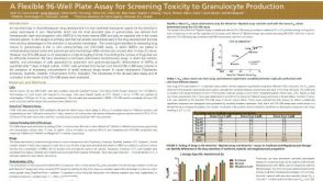 科学海报A Flexible 96-Well Plate Assay for Screening Toxicity to Granulocyte Production
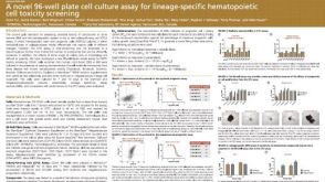 科学海报A Novel 96-well Plate Cell Culture Assay for Lineage-Specific Hematopoietic Cell Toxicity Screening
 沪公网安备31010102008431号