Cai S et al. (APR 2005)
Cancer research 65 8 3319--27
Mitochondrial targeting of human O6-methylguanine DNA methyltransferase protects against cell killing by chemotherapeutic alkylating agents.
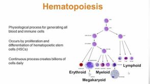
DNA repair capacity of eukaryotic cells has been studied extensively in recent years. Mammalian cells have been engineered to overexpress recombinant nuclear DNA repair proteins from ectopic genes to assess the impact of increased DNA repair capacity on genome stability. This approach has been used in this study to specifically target O(6)-methylguanine DNA methyltransferase (MGMT) to the mitochondria and examine its impact on cell survival after exposure to DNA alkylating agents. Survival of human hematopoietic cell lines and primary hematopoietic CD34(+) committed progenitor cells was monitored because the baseline repair capacity for alkylation-induced DNA damage is typically low due to insufficient expression of MGMT. Increased DNA repair capacity was observed when K562 cells were transfected with nuclear-targeted MGMT (nucl-MGMT) or mitochondrial-targeted MGMT (mito-MGMT). Furthermore,overexpression of mito-MGMT provided greater resistance to cell killing by 1,3-bis (2-chloroethyl)-1-nitrosourea (BCNU) than overexpression of nucl-MGMT. Simultaneous overexpression of mito-MGMT and nucl-MGMT did not enhance the resistance provided by mito-MGMT alone. Overexpression of either mito-MGMT or nucl-MGMT also conferred a similar level of resistance to methyl methanesulfonate (MMS) and temozolomide (TMZ) but simultaneous overexpression in both cellular compartments was neither additive nor synergistic. When human CD34(+) cells were infected with oncoretroviral vectors that targeted O(6)-benzylguanine (6BG)-resistant MGMT (MGMT(P140K)) to the nucleus or the mitochondria,committed progenitors derived from infected cells were resistant to 6BG/BCNU or 6BG/TMZ. These studies indicate that mitochondrial or nuclear targeting of MGMT protects hematopoietic cells against cell killing by BCNU,TMZ,and MMS,which is consistent with the possibility that mitochondrial DNA damage and nuclear DNA damage contribute equally to alkylating agent-induced cell killing during chemotherapy.
View Publication
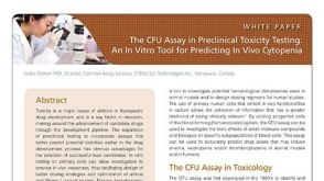
Scoring CFU-GM colonies in vitro by data fusion: a first account.
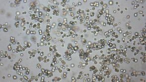
OBJECTIVE: In vitro models of hematopoiesis used in investigative hematopathology and in safety studies on candidate drugs,involve clonogenic assays on colony-forming unit granulocyte macrophage (CFU-GM). These assays require live and unstained colonies to be counted. Most laboratories still rely on visual scoring,which is time-consuming and error-prone. As a consequence,automated scoring is highly desired. An algorithm that recognizes and scores CFU-GM colonies by data fusion has been developed. Some preliminary results are presented in this article. METHODS: CFU-GM assays were carried out on hematopoietic progenitors (human umbilical cord blood cells) grown in methylcellulose. Colony images were acquired by a digital camera and stored. RESULTS: The classifier was designed to process images of layers sampled from a three-dimensional (3D) domain and forming a stack. Structure and texture information was extracted from each image. Classifier training was based on a 3D colony model applied to the image stack. The number of scored colonies (assigned class) was required to match the count supplied by the human expert (class of belonging). The trained classifier was validated on one more stack and then applied to a stack with overlapping colonies. Scoring in distortion- and caustic-affected border areas was also successfully demonstrated. Because of hardware limitations,compact colonies in some cases were missed. CONCLUSIONS: The industry's scoring methods all rely on structure alone and process 2D data. Instead,the classifier here fuses data from a whole stack and is capable,in principle,of high-throughput screening.
View Publication
产品号#:
产品名:
Fenouille N et al. (DEC 2010)
Cancer research 70 23 9659--70
Persistent activation of the Fyn/ERK kinase signaling axis mediates imatinib resistance in chronic myelogenous leukemia cells through upregulation of intracellular SPARC.
SPARC is an extracellular matrix protein that exerts pleiotropic effects on extracellular matrix organization,growth factor availability,cell adhesion,differentiation,and immunity in cancer. Chronic myelogenous leukemia (CML) cells resistant to the BCR-ABL inhibitor imatinib (IM-R cells) were found to overexpress SPARC mRNA. In this study,we show that imatinib triggers SPARC accumulation in a variety of tyrosine kinase inhibitor (TKI)-resistant CML cell lines. SPARC silencing in IM-R cells restored imatinib sensitivity,whereas enforced SPARC expression in imatinib-sensitive cells promoted viability as well as protection against imatinib-mediated apoptosis. Notably,we found that the protective effect of SPARC required intracellular retention inside cells. Accordingly,SPARC was not secreted into the culture medium of IM-R cells. Increased SPARC expression was intimately linked to persistent activation of the Fyn/ERK kinase signaling axis. Pharmacologic inhibition of this pathway or siRNA-mediated knockdown of Fyn kinase resensitized IM-R cells to imatinib. In support of our findings,increased levels of SPARC mRNA were documented in blood cells from CML patients after 1 year of imatinib therapy compared with initial diagnosis. Taken together,our results highlight an important role for the Fyn/ERK signaling pathway in imatinib-resistant cells that is driven by accumulation of intracellular SPARC.
View Publication
产品号#:
04100
产品名:
MethoCult™ H4100
Zhou L et al. (FEB 2011)
Cancer research 71 3 955--63
Reduced SMAD7 leads to overactivation of TGF-beta signaling in MDS that can be reversed by a specific inhibitor of TGF-beta receptor I kinase.
Even though myelodysplastic syndromes (MDS) are characterized by ineffective hematopoiesis,the molecular alterations that lead to marrow failure have not been well elucidated. We have previously shown that the myelosuppressive TGF-β pathway is constitutively activated in MDS progenitors. Because there is conflicting data about upregulation of extracellular TGF-β levels in MDS,we wanted to determine the molecular basis of TGF-β pathway overactivation and consequent hematopoietic suppression in this disease. We observed that SMAD7,a negative regulator of TGF-β receptor I (TBRI) kinase,is markedly decreased in a large meta-analysis of gene expression studies from MDS marrow-derived CD34(+) cells. SMAD7 protein was also found to be significantly decreased in MDS marrow progenitors when examined immunohistochemically in a bone marrow tissue microarray. Reduced expression of SMAD7 in hematopoietic cells led to increased TGF-β-mediated gene transcription and enhanced sensitivity to TGF-β-mediated suppressive effects. The increased TGF-β signaling due to SMAD7 reduction could be effectively inhibited by a novel clinically relevant TBRI (ALK5 kinase) inhibitor,LY-2157299. LY-2157299 could inhibit TGF-β-mediated SMAD2 activation and hematopoietic suppression in primary hematopoietic stem cells. Furthermore,in vivo administration of LY-2157299 ameliorated anemia in a TGF-β overexpressing transgenic mouse model of bone marrow failure. Most importantly,treatment with LY-2157199 stimulated hematopoiesis from primary MDS bone marrow specimens. These studies demonstrate that reduction in SMAD7 is a novel molecular alteration in MDS that leads to ineffective hematopoiesis by activating of TGF-β signaling in hematopoietic cells. These studies also illustrate the therapeutic potential of TBRI inhibitors in MDS.
View Publication
产品号#:
09600
09650
09850
产品名:
StemSpan™ SFEM
StemSpan™ SFEM
Kurtzberg LS et al. (MAY 2011)
Clinical cancer research : an official journal of the American Association for Cancer Research 17 9 2777--87
Genz-644282, a novel non-camptothecin topoisomerase I inhibitor for cancer treatment.
PURPOSE: Genz-644282 [8,9-dimethoxy-5-(2-N-methylaminoethyl)-2,3-methylenedioxy-5H-dibenzo[c,h][1,6]naphthyridin-6-one] has emerged as a promising candidate for antitumor agents. This report describes the bone marrow colony-forming unit,granulocyte macrophage (CFU-GM) and tumor cell CFU activity of topoisomerase I (Top1) inhibitors,such as Genz-644282,topotecan,irinotecan/SN-38,and ARC-111,and examines their activity in several human tumor xenograft models. EXPERIMENTAL DESIGN: Colony-forming assays were conducted with mouse and human bone marrow and eight human tumor cell lines. In addition,29 human tumor cell lines representing a range of histology and potential resistance mechanisms were assayed for sensitivity to Genz-644282 in a 72-hour exposure assay. The efficacy of Genz-644282 was compared with standard anticancer drugs (i.e.,irinotecan,docetaxel,and dacarbazine) in human tumor xenografts of colon cancer,renal cell carcinoma,non-small cell lung cancer,and melanoma. RESULTS: Human bone marrow CFU-GM was more sensitive to the Top1 inhibitors than was mouse bone marrow CFU-GM. The ratio of mouse to human IC(90) values was more than 10 for the camptothecins and less than 10 for Genz-644282,which had more potency as a cytotoxic agent toward human tumor cells in culture than the camptothecins in the colony-forming and 72-hour proliferation assays. Genz-644282 has superior or equal antitumor activity in the human tumor xenografts than the standard drug comparators. CONCLUSIONS: On the basis of preclinical activity and safety,Genz-644282 was selected for development and is currently undergoing phase 1 clinical trial.
View Publication
产品号#:
03434
03444
04035
84534
84544
产品名:
MethoCult™ GF M3434
MethoCult™ GF M3434
MethoCult™ 不含EPO的H4035 Optimum
MethoCult GF H84534, 100mL
Yang Y et al. (JUN 2011)
Experimental biology and medicine (Maywood,N.J.) 236 6 729--35
Protective effect of dammarane sapogenins against chemotherapy-induced myelosuppression in mice.
Chemotherapy is the most common way to treat malignancies,but myelosuppression,one of its common side-effects,is a formidable problem. The present study described the protective role of dammarane sapogenins (DS),an active fraction from oriental ginseng,on myelosuppression induced by cyclophosphamide (CP) in mice. DS was orally administered at different dosages (37.5,75,and 150 mg/kg) for 10 d after CP administration (200 mg/kg intraperitoneally). The results showed that DS increased the number of white blood cells (WBC) on day 3 and day 7 (P textless 0.05),such that WBC levels were increased by 105.7 ± 29.5% at 75 mg/kg of DS on day 3 (P textless 0.05,compared with the CP group). Similar results were observed in red blood cells and platelets in DS-treated groups. The colony-forming assay demonstrated that the depressed numbers of CFU-GM (colony-forming unit-granulocyte and macrophage),CFU-E (colony-forming unit-erythroid),BFU-E (burst-forming unit-erythroid),CFU-Meg (colony-forming unit-megakaryocyte) and CFU-GEMM (colony-forming unit-granulocyte,-erythrocyte,-monocyte and -megakaryocyte) induced by CP were significantly reversed after DS treatment. Moreover,the ameliorative effect of DS on myelosuppression was also observed in the femur by hematoxylin/eosin staining. In DS-treated groups,ConA-induced splenocyte proliferation was enhanced significantly at all the doses (37.5,75,150 mg/kg) on day 3 at the rate of 50.3 ± 8.0%,77.6 ± 8.5% and 44.5 ± 8.4%,respectively,while lipopolysaccharide-induced proliferation was increased mainly on day 7 (P textless 0.01),with an increased rate of 39.8 ± 5.6%,34.9 ± 6.6% and 38.3 ± 7.3%,respectively. The thymus index was also markedly increased by 70.4% and 36.6% at 75 mg/kg on days 3 and 7,respectively,as compared with the CP group. In summary,DS has a protective function against CP-induced myelosuppression. Its mechanism might be related to stimulating hematopoiesis recovery,as well as enhancing the immunological function.
View Publication
Pfeifer A et al. (SEP 2001)
Proceedings of the National Academy of Sciences of the United States of America 98 20 11450--5
Delivery of the Cre recombinase by a self-deleting lentiviral vector: efficient gene targeting in vivo.
The Cre recombinase (Cre) from bacteriophage P1 is an important tool for genetic engineering in mammalian cells. We constructed lentiviral vectors that efficiently deliver Cre in vitro and in vivo. Surprisingly,we found a significant reduction in proliferation and an accumulation in the G(2)/M phase of Cre-expressing cells. To minimize the toxic effect of Cre,we designed a lentiviral vector that integrates into the host genome,expresses Cre in the target cell,and is subsequently deleted from the genome in a Cre-dependent manner. Thus,the activity of Cre terminates its own expression (self-deleting). We showed efficient modification of target genes in vitro and in the brain after transduction with the self-deleting vectors. In contrast to sustained Cre expression,transient expression of Cre from the self-deleting vector induced significantly less cytotoxicity. Such a self-deleting Cre vector is a promising tool for the induction of conditional gene modifications with minimal Cre toxicity in vivo.
View Publication

 EasySep™小鼠TIL(CD45)正选试剂盒


 沪公网安备31010102008431号