
 EasySep™小鼠TIL(CD45)正选试剂盒


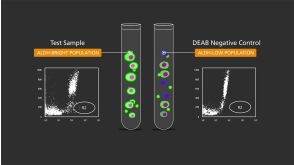
技术资料
-
 技术公告Generation of Natural Killer Cells from Human Pluripotent Stem Cells Using STEMdiff™ and StemSpan™ Media and Supplements
细胞类型:
NK细胞,多能干细胞
发布日期: 07/30/2021 -
 31:39
线上讲座Serum- and Feeder-Free Differentiation of Erythroid Progenitor Cells from hPSCs发布日期: 05/21/2021
31:39
线上讲座Serum- and Feeder-Free Differentiation of Erythroid Progenitor Cells from hPSCs发布日期: 05/21/2021 -
 挂图Derivation and Applications of Human Pluripotent Stem Cells Overview of the derivation of human embryonic stem cells (hESCs) and induced pluripotent stem cells (iPSCs)发布日期: 11/26/2020
 -
 1:02:03
线上讲座Building Brain Organoids and AssemBloids™ to Study Human Development and Disease发布日期: 10/30/2020
1:02:03
线上讲座Building Brain Organoids and AssemBloids™ to Study Human Development and Disease发布日期: 10/30/2020


 沪公网安备31010102008431号