Inhibition of osteoclast function reduces hematopoietic stem cell numbers in vivo.
Osteoblasts play a crucial role in the hematopoietic stem cell (HSC) niche; however,an overall increase in their number does not necessarily promote hematopoiesis. Because the activity of osteoblasts and osteoclasts is coordinately regulated,we hypothesized that active bone-resorbing osteoclasts would participate in HSC niche maintenance. Mice treated with bisphosphonates exhibited a decrease in proportion and absolute number of Lin(-)cKit(+)Sca1(+) Flk2(-) (LKS Flk2(-)) and long-term culture-initiating cells in bone marrow (BM). In competitive transplantation assays,the engraftment of treated BM cells was inferior to that of controls,confirming a decrease in HSC numbers. Accordingly,bisphosphonates abolished the HSC increment produced by parathyroid hormone. In contrast,the number of colony-forming-unit cells in BM was increased. Because a larger fraction of LKS in the BM of treated mice was found in the S/M phase of the cell cycle,osteoclast impairment makes a proportion of HSCs enter the cell cycle and differentiate. To prove that HSC impairment was a consequence of niche manipulation,a group of mice was treated with bisphosphonates and then subjected to BM transplantation from untreated donors. Treated recipient mice experienced a delayed hematopoietic recovery compared with untreated controls. Our findings demonstrate that osteoclast function is fundamental in the HSC niche.
View Publication
产品号#:
03434
03444
05350
产品名:
MethoCult™ GF M3434
MethoCult™ GF M3434
Li J et al. (MAR 2005)
Clinical Cancer Research 11 6 2195--2204
Generation of PRL-3- and PRL-1-specific monoclonal antibodies as potential diagnostic markers for cancer metastases
PURPOSE: The PRL-3 mRNA is consistently elevated in metastatic samples derived from colorectal cancers. We sought to generate a specific PRL-3 monoclonal antibody (mAb) that might serve as a potential diagnostic marker for colorectal cancer metastasis. EXPERIMENTAL DESIGN: PRL-3 is one of three members (PRL-1,PRL-2,and PRL-3) in a unique protein-tyrosine phosphatase family. Because the three PRLs are 76% to 87% identical in their amino acid sequences,it poses a great challenge to obtain mAbs that are specific for respective phosphatase of regenerating liver (PRL) but not for the other two in the family. We screened over 1,400 hybridoma clones to generate mAbs specific to each PRL member. RESULTS: We obtained two hybridoma clones specifically against PRL-3 and another two clones specifically against PRL-1. These antibodies had been evaluated by several critical tests to show their own specificities and applications. Most importantly,the PRL-3 mAbs were assessed on 282 human colorectal tissue samples (121 normal,17 adenomas,and 144 adenocarcinomas). PRL-3 protein was detected in 11% of adenocarcinoma samples. The PRL-3- and PRL-1-specific mAbs were further examined on 204 human multiple cancer tissues. The differential expressions of PRL-3 and PRL-1 confirmed the mAbs' specificity. CONCLUSIONS: Using several approaches,we show that PRL-3- or PRL-1-specific mAbs react only to their respective antigen. The expression of PRL-3 in textgreater10% of primary colorectal cancer samples indicates that PRL-3 may prime the metastatic process. These mAbs will be useful as markers in clinical diagnosis for assessing tumor aggressiveness.
View Publication
Vieillard V et al. (AUG 2005)
Proceedings of the National Academy of Sciences 102 31 10981--86
NK cytotoxicity against CD4+ T cells during HIV-1 infection: A gp41 peptide induces the expression of an NKp44 ligand
HIV infection leads to a state of chronic immune activation and progressive deterioration in immune function,manifested most recognizably by the progressive depletion of CD4+ T cells. A substantial percentage of natural killer (NK) cells from patients with HIV infection are activated and express the natural cytotoxicity receptor (NCR) NKp44. Here we show that a cellular ligand for NKp44 (NKp44L) is expressed during HIV-1 infection and is correlated with both the progression of CD4+ T cell depletion and the increase of viral load. CD4+ T cells expressing this ligand are highly sensitive to the NK lysis activity mediated by NKp44+ NK cells. The expression of NKp44L is induced by the linear motif NH2-SWSNKS-COOH of the HIV-1 envelope gp41 protein. This highly conserved motif appears critical to the sharp increase in NK lysis of CD4+ T cells from HIV-infected patients. These studies strongly suggest that induction of NKp44L plays a key role in the lysis of CD4+ T cells by activated NK cells in HIV infection and consequently provide a framework for considering how HIV-1 may use NK cell immune surveillance to trigger CD4+ T cells. Understanding this mechanism may help to develop future therapeutic strategies and vaccines against HIV-1 infection.
View Publication
产品号#:
03800
03801
03802
03803
03804
03805
03806
05150
15021
15061
产品名:
ClonaCell™-HY杂交瘤试剂盒
ClonaCell™-HY培养基A
ClonaCell™-HY 培养基 B
ClonaCell™-HY 培养基 C
ClonaCell™-HY 培养基 D
ClonaCell™-HY 培养基 E
ClonaCell™-HY PEG
MyeloCult™ H5100
RosetteSep™人T细胞富集抗体混合物
RosetteSep™人T细胞富集抗体混合物
Crook JM et al. (MAR 2015)
Expert review of neurotherapeutics 15 3 295--304
The potential of induced pluripotent stem cells in models of neurological disorders: implications on future therapy.
There is an urgent need for new and advanced approaches to modeling the pathological mechanisms of complex human neurological disorders. This is underscored by the decline in pharmaceutical research and development efficiency resulting in a relative decrease in new drug launches in the last several decades. Induced pluripotent stem cells represent a new tool to overcome many of the shortcomings of conventional methods,enabling live human neural cell modeling of complex conditions relating to aberrant neurodevelopment,such as schizophrenia,epilepsy and autism as well as age-associated neurodegeneration. This review considers the current status of induced pluripotent stem cell-based modeling of neurological disorders,canvassing proven and putative advantages,current constraints,and future prospects of next-generation culture systems for biomedical research and translation.
View Publication
产品号#:
05850
05857
05870
05875
85850
85857
85870
85875
产品名:
mTeSR™1
mTeSR™1
Easley CA et al. (MAY 2015)
Stem Cell Research 14 3 347--355
Assessing reproductive toxicity of two environmental toxicants with a novel in vitro human spermatogenic model
Environmental influences and insults by reproductive toxicant exposure can lead to impaired spermatogenesis or infertility. Understanding how toxicants disrupt spermatogenesis is critical for determining how environmental factors contribute to impaired fertility. While current animal models are available,understanding of the reproductive toxic effects on human fertility requires a more robust model system. We recently demonstrated that human pluripotent stem cells can differentiate into spermatogonial stem cells/spermatogonia,primary and secondary spermatocytes,and haploid spermatids; a model that mimics many aspects of human spermatogenesis. Here,using this model system,we examine the effects of 2-bromopropane (2-BP) and 1,2,dibromo-3-chloropropane (DBCP) on in vitro human spermatogenesis. 2-BP and DBCP are non-endocrine disrupting toxicants that are known to impact male fertility. We show that acute treatment with either 2-BP or DBCP induces a reduction in germ cell viability through apoptosis. 2-BP and DBCP affect viability of different cell populations as 2-BP primarily reduces spermatocyte viability,whereas DBCP exerts a much greater effect on spermatogonia. Acute treatment with 2-BP or DBCP also reduces the percentage of haploid spermatids. Both 2-BP and DBCP induce reactive oxygen species (ROS) formation leading to an oxidized cellular environment. Taken together,these results suggest that acute exposure with 2-BP or DBCP causes human germ cell death in vitro by inducing ROS formation. This system represents a unique platform for assessing human reproductive toxicity potential of various environmental toxicants in a rapid,efficient,and unbiased format.
View Publication
产品号#:
05850
05857
05870
05875
85850
85857
85870
85875
产品名:
mTeSR™1
mTeSR™1
Feng R et al. (MAR 2007)
Blood 109 5 2130--8
SDX-308, a nonsteroidal anti-inflammatory agent, inhibits NF-kappaB activity, resulting in strong inhibition of osteoclast formation/activity and multiple myeloma cell growth.
Multiple myeloma is characterized by increased osteoclast activity that results in bone destruction and lytic lesions. With the prolonged overall patient survival achieved by new treatment modalities,additional drugs are required to inhibit bone destruction. We focused on a novel and more potent structural analog of the nonsteroidal anti-inflammatory drug etodolac,known as SDX-308,and its effects on osteoclastogenesis and multiple myeloma cells. SDX-101 is another structural analog of etodolac that is already used in clinical trials for the treatment of B-cell chronic lymphocytic leukemia (B-CLL). Compared with SDX-101,a 10-fold lower concentration of SDX-308 induced potent (60%-80%) inhibition of osteoclast formation,and a 10- to 100-fold lower concentration inhibited multiple myeloma cell proliferation. Bone resorption was completely inhibited by SDX-308,as determined in dentin-based bone resorption assays. SDX-308 decreased constitutive and RANKL-stimulated NF-kappaB activation and osteoclast formation in an osteoclast cellular model,RAW 264.7. SDX-308 effectively suppressed TNF-alpha-induced IKK-gamma and IkappaB-alpha phosphorylation and degradation and subsequent NF-kappaB activation in human multiple myeloma cells. These results indicate that SDX-308 effectively inhibits multiple myeloma cell proliferation and osteoclast activity,potentially by controlling NF-kappaB activation signaling. We propose that SDX-308 is a promising therapeutic candidate to inhibit multiple myeloma growth and osteoclast activity and that it should receive attention for further study.
View Publication
产品号#:
04434
04444
产品名:
MethoCult™ H4434 Classic
MethoCult™ H4434 Classic
Ling SSM et al. (JUN 2015)
PLOS ONE 10 6 e0131460
Instrumental Role of Helicobacter pylori γ-Glutamyl Transpeptidase in VacA-Dependent Vacuolation in Gastric Epithelial Cells
Helicobacter pylori causes cellular vacuolation in host cells,a cytotoxic event attributed to vacuolating cytotoxin (VacA) and the presence of permeant weak bases such as ammonia. We report here the role of γ-glutamyl transpeptidase (GGT),a constitutively expressed secretory enzyme of H. pylori,in potentiating VacA-dependent vacuolation formation in H. pylori-infected AGS and primary gastric cells. The enhancement is brought about by GGT hydrolysing glutamine present in the extracellular medium,thereby releasing ammonia which accentuates the VacA-induced vacuolation. The events of vacuolation in H. pylori wild type (WT)- and Δggt-infected AGS cells were first captured and visualized by real-time phase-contrast microscopy where WT was observed to induce more vacuoles than Δggt. By using semi-quantitative neutral red uptake assay,we next showed that Δggt induced significantly less vacuolation in AGS and primary gastric epithelial cells as compared to the parental strain (Ptextless0.05) indicating that GGT potentiates the vacuolating effect of VacA. Notably,vacuolation induced by WT was significantly reduced in the absence of GGT substrate,glutamine (Ptextless0.05) or in the presence of a competitive GGT inhibitor,serine-borate complex. Furthermore,the vacuolating ability of Δggt was markedly restored when co-incubated with purified recombinant GGT (rGGT),although rGGT itself did not induce vacuolation independently. Similarly,the addition of exogenous ammonium chloride as a source of ammonia also rescued the ability of Δggt to induce vacuolation. Additionally,we also show that monoclonal antibodies against GGT effectively inhibited GGT activity and successfully suppressed H. pylori-induced vacuolation. Collectively,our results clearly demonstrate that generation of ammonia by GGT through glutamine hydrolysis is responsible for enhancing VacA-dependent vacuolation. Our findings provide a new perspective on GGT as an important virulence factor and a promising target in the management of H. pylori-associated gastric diseases.
View Publication
产品号#:
03800
03801
03802
03803
03804
03805
03806
产品名:
ClonaCell™-HY杂交瘤试剂盒
ClonaCell™-HY培养基A
ClonaCell™-HY 培养基 B
ClonaCell™-HY 培养基 C
ClonaCell™-HY 培养基 D
ClonaCell™-HY 培养基 E
ClonaCell™-HY PEG
Kia R et al. (MAR 2015)
Toxicological Sciences 144 1 173--185
MicroRNA-122: a novel hepatocyte-enriched in vitro marker of drug-induced cellular toxicity.
Emerging hepatic models for the study of drug-induced toxicity include pluripotent stem cell-derived hepatocyte-like cells (HLCs) and complex hepatocyte-non-parenchymal cellular coculture to mimic the complex multicellular interactions that recapitulate the niche environment in the human liver. However,a specific marker of hepatocyte perturbation,required to discriminate hepatocyte damage from non-specific cellular toxicity contributed by non-hepatocyte cell types or immature differentiated cells is currently lacking,as the cytotoxicity assays routinely used in in vitro toxicology research depend on intracellular molecules which are ubiquitously present in all eukaryotic cell types. In this study,we demonstrate that microRNA-122 (miR-122) detection in cell culture media can be used as a hepatocyte-enriched in vitro marker of drug-induced toxicity in homogeneous cultures of hepatic cells,and a cell-specific marker of toxicity of hepatic cells in heterogeneous cultures such as HLCs generated from various differentiation protocols and pluripotent stem cell lines,where conventional cytotoxicity assays using generic cellular markers may not be appropriate. We show that the sensitivity of the miR-122 cytotoxicity assay is similar to conventional assays that measure lactate dehydrogenase activity and intracellular adenosine triphosphate when applied in hepatic models with high levels of intracellular miR-122,and can be multiplexed with other assays. MiR-122 as a biomarker also has the potential to bridge results in in vitro experiments to in vivo animal models and human samples using the same assay,and to link findings from clinical studies in determining the relevance of in vitro models being developed for the study of drug-induced liver injury.
View Publication

 EasySep™小鼠TIL(CD45)正选试剂盒


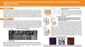 科学海报Expansion and Differentiation Potential of Human Pluripotent Stem Cells Maintained in cGMP Animal Origin-Free Medium
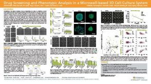 科学海报Drug Screening and Phenotypic Analysis in a Microwell-based 3D Cell Culture System


 沪公网安备31010102008431号