Shi S et al. (SEP 2011)
Journal of Visualized Experiments 55 e3010
A high-throughput automated platform for the development of manufacturing cell lines for protein therapeutics
The fast-growing biopharmaceutical industry demands speedy development of highly efficient and reliable production systems to meet the increasing requirement for drug supplies. The generation of production cell lines has traditionally involved manual operations that are labor-intensive,low-throughput and vulnerable to human errors. We report here an integrated high-throughput and automated platform for development of manufacturing cell lines for the production of protein therapeutics. The combination of BD FACS Aria Cell Sorter,CloneSelect Imager and TECAN Freedom EVO liquid handling system has enabled a high-throughput and more efficient cell line development process. In this operation,production host cells are first transfected with an expression vector carrying the gene of interest (1),followed by the treatment with a selection agent. The stably-transfected cells are then stained with fluorescence-labeled anti-human IgG antibody,and are subsequently subject to flow cytometry analysis (2-4). Highly productive cells are selected based on fluorescence intensity and are isolated by single-cell sorting on a BD FACSAria. Colony formation from single-cell stage was detected microscopically and a series of time-laps digital images are taken by CloneSelect Imager for the documentation of cell line history. After single clones have formed,these clones were screened for productivity by ELISA performed on a TECAN Freedom EVO liquid handling system. Approximately 2,000 - 10,000 clones can be screened per operation cycle with the current system setup. This integrated approach has been used to generate high producing Chinese hamster ovary (CHO) cell lines for the production of therapeutic monoclonal antibody (mAb) as well as their fusion proteins. With the aid of different types of detecting probes,the method can be used for developing other protein therapeutics or be applied to other production host systems. Comparing to the traditional manual procedure,this automated platform demonstrated advantages of significantly increased capacity,ensured clonality,traceability in cell line history with electronic documentation and much reduced opportunity in operator error.
View Publication
产品号#:
30000
产品名:
Smith Sa et al. (MAR 2012)
Journal of Virology 86 5 2665--75
Persistence of circulating memory B cell clones with potential for Dengue virus disease enhancement for decades following infection
Symptomatic dengue virus infection ranges in disease severity from an influenza-like illness to life-threatening shock. One model of the mechanism underlying severe disease proposes that weakly neutralizing,dengue serotype cross-reactive antibodies induced during a primary infection facilitate virus entry into Fc receptor-bearing cells during a subsequent secondary infection,increasing viral replication and the release of cytokines and vasoactive mediators,culminating in shock. This process has been termed antibody-dependent enhancement of infection and has significantly hindered vaccine development. Much of our understanding of this process has come from studies using mouse monoclonal antibodies (MAbs); however,antibody responses in mice typically exhibit less complexity than those in humans. A better understanding of the humoral immune response to natural dengue virus infection in humans is sorely needed. Using a high-efficiency human hybridoma technology,we isolated 37 hybridomas secreting human MAbs to dengue viruses from 12 subjects years or even decades following primary or secondary infection. The majority of the human antibodies recovered were broadly cross-reactive,directed against either envelope or premembrane proteins,and capable of enhancement of infection in vitro; few exhibited serotype-specific binding or potent neutralizing activity. Memory B cells encoding enhancing antibodies predominated in the circulation,even two or more decades following infection. Mapping the epitopes and activity of naturally occurring dengue antibodies should prove valuable in determining whether the enhancing and neutralizing activity of antibodies can be separated. Such principles could be used in the rational design of vaccines that enhance the induction of neutralizing antibodies,while lowering the risk of dengue shock syndrome.
View Publication
产品号#:
03800
03801
03802
03803
03804
03805
03806
产品名:
ClonaCell™-HY杂交瘤试剂盒
ClonaCell™-HY培养基A
ClonaCell™-HY 培养基 B
ClonaCell™-HY 培养基 C
ClonaCell™-HY 培养基 D
ClonaCell™-HY 培养基 E
ClonaCell™-HY PEG
Cabral TM et al. (JUL 2012)
Journal of Virological Methods 183 1 25--33
Development and characterization of neutralizing monoclonal antibodies against the pandemic H1N1 virus (2009).
The 2009 H1N1 influenza pandemic was a major international public health crisis which caused considerable morbidity and mortality worldwide. The goal of this study was to produce anti-H1 monoclonal antibodies (MAbs) for improving diagnostic immunological assays and to develop potential immunotherapeutics. Nine MAbs were produced after immunizing mice with recombinant hemagglutinin (HA) protein from A/California/06/09. Two spleenocyte myeloma fusions yielded 1588 hybridoma cultures. After screening the hybridoma culture supernatants for antibody reactivity to rHA,nine clones were selected for further characterization. Cross-reactivity studies of the anti-rHA antibodies against a panel of influenza viruses (H1-H16) revealed eight out of nine MAbs were specific to the pandemic H1 subtype,except for MAb F256G2sc1 which also cross-reacted with H5 subtype virus. All MAbs were of the IgG1κ isotype,except F256G2sc1 which was IgG2aκ. The anti-rHA MAbs had binding affinities to rHA that ranged from a K(D) (disassociation constant) of 1.34×10(-9)M (F255G7sc1) to the weakest affinity of 4.60×10(-8)M (F255G4sc1). Interestingly,in a plaque reduction neutralization assay,all MAbs except F255G3sc1 demonstrated neutralizing ability. Furthermore,all MAbs except F255G3sc1 and F255G9sc1 exhibited anti-hemagglutinin activity against pandemic H1N1 viruses,but not against classical North American swine influenza viruses of the same subtype. Immunofluorescence assay (IFA) demonstrated that all MAbs except F255G1sc1 and F255G3sc1 were able to detect 2009 pandemic H1N1 (2009) virus- infected MDCK cells. The MAbs were also evaluated for potential use in competitive ELISA (cELISA),and with the exception of F255G3sc1,all MAbs showed competitive activity with serum collected from pigs infected with pandemic H1N1 virus (2009). The developed MAbs have demonstrated utility as immunodiagnostic and research reagents,and their neutralizing capabilities also hold potential for designing antiviral drugs against pandemic influenza.
View Publication
产品号#:
03800
03801
03802
03803
03804
03805
03806
产品名:
ClonaCell™-HY杂交瘤试剂盒
ClonaCell™-HY培养基A
ClonaCell™-HY 培养基 B
ClonaCell™-HY 培养基 C
ClonaCell™-HY 培养基 D
ClonaCell™-HY 培养基 E
ClonaCell™-HY PEG
Xu H et al. (JUL 2016)
Organic & biomolecular chemistry 14 26 6179--83
Cellular thermal shift and clickable chemical probe assays for the determination of drug-target engagement in live cells.
Proof of drug-target engagement in physiologically-relevant contexts is a key pillar of successful therapeutic target validation. We developed two orthogonal technologies,the cellular thermal shift assay (CETSA) and a covalent chemical probe reporter approach (harnessing sulfonyl fluoride tyrosine labeling and subsequent click chemistry) to measure the occupancy of the mRNA-decapping scavenger enzyme DcpS by a small molecule inhibitor in live cells. Enzyme affinity determined using isothermal dose response fingerprinting (ITDRFCETSA) and the concentration required to occupy 50% of the enzyme (OC50) using the chemical probe reporter assay were very similar. In this case,the chemical probe method worked well due to the long offset kinetics of the reversible inhibitor (determined using a fluorescent dye-tagged probe). This work suggests that CETSA could become the first choice assay to determine in-cell target engagement due to its simplicity.
View Publication
Scoring CFU-GM colonies in vitro by data fusion: a first account.
OBJECTIVE: In vitro models of hematopoiesis used in investigative hematopathology and in safety studies on candidate drugs,involve clonogenic assays on colony-forming unit granulocyte macrophage (CFU-GM). These assays require live and unstained colonies to be counted. Most laboratories still rely on visual scoring,which is time-consuming and error-prone. As a consequence,automated scoring is highly desired. An algorithm that recognizes and scores CFU-GM colonies by data fusion has been developed. Some preliminary results are presented in this article. METHODS: CFU-GM assays were carried out on hematopoietic progenitors (human umbilical cord blood cells) grown in methylcellulose. Colony images were acquired by a digital camera and stored. RESULTS: The classifier was designed to process images of layers sampled from a three-dimensional (3D) domain and forming a stack. Structure and texture information was extracted from each image. Classifier training was based on a 3D colony model applied to the image stack. The number of scored colonies (assigned class) was required to match the count supplied by the human expert (class of belonging). The trained classifier was validated on one more stack and then applied to a stack with overlapping colonies. Scoring in distortion- and caustic-affected border areas was also successfully demonstrated. Because of hardware limitations,compact colonies in some cases were missed. CONCLUSIONS: The industry's scoring methods all rely on structure alone and process 2D data. Instead,the classifier here fuses data from a whole stack and is capable,in principle,of high-throughput screening.
View Publication
产品号#:
产品名:
Fenouille N et al. (DEC 2010)
Cancer research 70 23 9659--70
Persistent activation of the Fyn/ERK kinase signaling axis mediates imatinib resistance in chronic myelogenous leukemia cells through upregulation of intracellular SPARC.
SPARC is an extracellular matrix protein that exerts pleiotropic effects on extracellular matrix organization,growth factor availability,cell adhesion,differentiation,and immunity in cancer. Chronic myelogenous leukemia (CML) cells resistant to the BCR-ABL inhibitor imatinib (IM-R cells) were found to overexpress SPARC mRNA. In this study,we show that imatinib triggers SPARC accumulation in a variety of tyrosine kinase inhibitor (TKI)-resistant CML cell lines. SPARC silencing in IM-R cells restored imatinib sensitivity,whereas enforced SPARC expression in imatinib-sensitive cells promoted viability as well as protection against imatinib-mediated apoptosis. Notably,we found that the protective effect of SPARC required intracellular retention inside cells. Accordingly,SPARC was not secreted into the culture medium of IM-R cells. Increased SPARC expression was intimately linked to persistent activation of the Fyn/ERK kinase signaling axis. Pharmacologic inhibition of this pathway or siRNA-mediated knockdown of Fyn kinase resensitized IM-R cells to imatinib. In support of our findings,increased levels of SPARC mRNA were documented in blood cells from CML patients after 1 year of imatinib therapy compared with initial diagnosis. Taken together,our results highlight an important role for the Fyn/ERK signaling pathway in imatinib-resistant cells that is driven by accumulation of intracellular SPARC.
View Publication
Zhou L et al. (FEB 2011)
Cancer research 71 3 955--63
Reduced SMAD7 leads to overactivation of TGF-beta signaling in MDS that can be reversed by a specific inhibitor of TGF-beta receptor I kinase.
Even though myelodysplastic syndromes (MDS) are characterized by ineffective hematopoiesis,the molecular alterations that lead to marrow failure have not been well elucidated. We have previously shown that the myelosuppressive TGF-β pathway is constitutively activated in MDS progenitors. Because there is conflicting data about upregulation of extracellular TGF-β levels in MDS,we wanted to determine the molecular basis of TGF-β pathway overactivation and consequent hematopoietic suppression in this disease. We observed that SMAD7,a negative regulator of TGF-β receptor I (TBRI) kinase,is markedly decreased in a large meta-analysis of gene expression studies from MDS marrow-derived CD34(+) cells. SMAD7 protein was also found to be significantly decreased in MDS marrow progenitors when examined immunohistochemically in a bone marrow tissue microarray. Reduced expression of SMAD7 in hematopoietic cells led to increased TGF-β-mediated gene transcription and enhanced sensitivity to TGF-β-mediated suppressive effects. The increased TGF-β signaling due to SMAD7 reduction could be effectively inhibited by a novel clinically relevant TBRI (ALK5 kinase) inhibitor,LY-2157299. LY-2157299 could inhibit TGF-β-mediated SMAD2 activation and hematopoietic suppression in primary hematopoietic stem cells. Furthermore,in vivo administration of LY-2157299 ameliorated anemia in a TGF-β overexpressing transgenic mouse model of bone marrow failure. Most importantly,treatment with LY-2157199 stimulated hematopoiesis from primary MDS bone marrow specimens. These studies demonstrate that reduction in SMAD7 is a novel molecular alteration in MDS that leads to ineffective hematopoiesis by activating of TGF-β signaling in hematopoietic cells. These studies also illustrate the therapeutic potential of TBRI inhibitors in MDS.
View Publication
产品号#:
09600
09650
09850
产品名:
StemSpan™ SFEM
StemSpan™ SFEM
Gu Z et al. (FEB 2006)
Antimicrobial agents and chemotherapy 50 2 625--31
In vitro antiretroviral activity and in vitro toxicity profile of SPD754, a new deoxycytidine nucleoside reverse transcriptase inhibitor for treatment of human immunodeficiency virus infection.
SPD754 (AVX754) is a deoxycytidine analogue nucleotide reverse transcriptase inhibitor (NRTI) in clinical development. These studies characterized the in vitro activity of SPD754 against NRTI-resistant human immunodeficiency virus type 1 (HIV-1) and non-clade B HIV-1 isolates,its activity in combination with other antiretrovirals,and its potential myelotoxicity and mitochondrial toxicity. SPD754 was tested against 50 clinical HIV-1 isolates (5 wild-type isolates and 45 NRTI-resistant isolates) in MT-4 cells using the Antivirogram assay. SPD754 susceptibility was reduced 1.2- to 2.2-fold against isolates resistant to zidovudine (M41L,T215Y/F,plus a median of three additional nucleoside analogue mutations [NAMs]) and/or lamivudine (M184V) and was reduced 1.3- to 2.8-fold against isolates resistant to abacavir (L74V,Y115F,and M184V plus one other NAM) or stavudine (V75T/M,M41L,T215F/Y,and four other NAMs). Insertions at amino acid position 69 and Q151M mutations (with or without M184V) reduced SPD754 susceptibility 5.2-fold and 14- to 16-fold,respectively (these changes gave values comparable to or less than the corresponding values for zidovudine,lamivudine,abacavir,and didanosine). SPD754 showed similar activity against isolates of group M HIV-1 clades,including A/G,B,C,D,A(E),D/F,F,and H. SPD754 showed additive effects in combination with other NRTIs,tenofovir,nevirapine,or saquinavir. SPD754 had no significant effects on cell viability or mitochondrial DNA in HepG2 or MT-4 cells during 28-day exposure at concentrations up to 200 microM. SPD754 showed a low potential for myelotoxicity against human bone marrow. In vitro,SPD754 retained activity against most NRTI-resistant HIV-1 clinical isolates and showed a low propensity to cause myelotoxicity and mitochondrial toxicity.
View Publication
产品号#:
04434
04444
产品名:
MethoCult™ H4434 Classic
MethoCult™ H4434 Classic
Cai S et al. (APR 2005)
Cancer research 65 8 3319--27
Mitochondrial targeting of human O6-methylguanine DNA methyltransferase protects against cell killing by chemotherapeutic alkylating agents.
DNA repair capacity of eukaryotic cells has been studied extensively in recent years. Mammalian cells have been engineered to overexpress recombinant nuclear DNA repair proteins from ectopic genes to assess the impact of increased DNA repair capacity on genome stability. This approach has been used in this study to specifically target O(6)-methylguanine DNA methyltransferase (MGMT) to the mitochondria and examine its impact on cell survival after exposure to DNA alkylating agents. Survival of human hematopoietic cell lines and primary hematopoietic CD34(+) committed progenitor cells was monitored because the baseline repair capacity for alkylation-induced DNA damage is typically low due to insufficient expression of MGMT. Increased DNA repair capacity was observed when K562 cells were transfected with nuclear-targeted MGMT (nucl-MGMT) or mitochondrial-targeted MGMT (mito-MGMT). Furthermore,overexpression of mito-MGMT provided greater resistance to cell killing by 1,3-bis (2-chloroethyl)-1-nitrosourea (BCNU) than overexpression of nucl-MGMT. Simultaneous overexpression of mito-MGMT and nucl-MGMT did not enhance the resistance provided by mito-MGMT alone. Overexpression of either mito-MGMT or nucl-MGMT also conferred a similar level of resistance to methyl methanesulfonate (MMS) and temozolomide (TMZ) but simultaneous overexpression in both cellular compartments was neither additive nor synergistic. When human CD34(+) cells were infected with oncoretroviral vectors that targeted O(6)-benzylguanine (6BG)-resistant MGMT (MGMT(P140K)) to the nucleus or the mitochondria,committed progenitors derived from infected cells were resistant to 6BG/BCNU or 6BG/TMZ. These studies indicate that mitochondrial or nuclear targeting of MGMT protects hematopoietic cells against cell killing by BCNU,TMZ,and MMS,which is consistent with the possibility that mitochondrial DNA damage and nuclear DNA damage contribute equally to alkylating agent-induced cell killing during chemotherapy.
View Publication

 EasySep™小鼠TIL(CD45)正选试剂盒


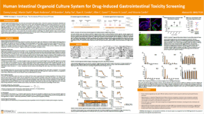 科学海报Human Intestinal Organoid Culture System for Drug-Induced Gastrointestinal Toxicity Screening
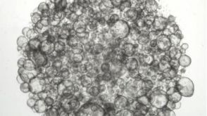
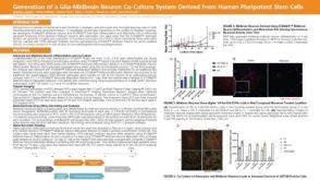 科学海报Generation of a Glia-Midbrain Neuron Co-Culture System Derived From Human Pluripotent Stem Cells
 沪公网安备31010102008431号