Microarray study reveals that HIV-1 induces rapid type-I interferon-dependent p53 mRNA up-regulation in human primary CD4+ T cells.
BACKGROUND: Infection with HIV-1 has been shown to alter expression of a large array of host cell genes. However,previous studies aimed at investigating the putative HIV-1-induced modulation of host gene expression have been mostly performed in established human cell lines. To better approximate natural conditions,we monitored gene expression changes in a cell population highly enriched in human primary CD4+ T lymphocytes exposed to HIV-1 using commercial oligonucleotide microarrays from Affymetrix. RESULTS: We report here that HIV-1 influences expression of genes related to many important biological processes such as DNA repair,cellular cycle,RNA metabolism and apoptosis. Notably,expression of the p53 tumor suppressor and genes involved in p53 homeostasis such as GADD34 were up-regulated by HIV-1 at the mRNA level. This observation is distinct from the previously reported p53 phosphorylation and stabilization at the protein level,which precedes HIV-1-induced apoptosis. We present evidence that the HIV-1-mediated increase in p53 gene expression is associated with virus-mediated induction of type-I interferon (i.e. IFN-alpha and IFN-beta). CONCLUSION: These observations have important implications for our understanding of HIV-1 pathogenesis,particularly in respect to the virus-induced depletion of CD4+ T cells.
View Publication
产品号#:
15022
15062
产品名:
RosetteSep™人CD4+ T细胞富集抗体混合物
RosetteSep™人CD4+ T细胞富集抗体混合物
Popovic R et al. (APR 2009)
Blood 113 14 3314--22
Regulation of mir-196b by MLL and its overexpression by MLL fusions contributes to immortalization.
Chromosomal translocations involving the Mixed Lineage Leukemia (MLL) gene produce chimeric proteins that cause abnormal expression of a subset of HOX genes and leukemia development. Here,we show that MLL normally regulates expression of mir-196b,a hematopoietic microRNA located within the HoxA cluster,in a pattern similar to that of the surrounding 5' Hox genes,Hoxa9 and Hoxa10,during embryonic stem (ES) cell differentiation. Within the hematopoietic lineage,mir-196b is most abundant in short-term hematopoietic stem cells and is down-regulated in more differentiated hematopoietic cells. Leukemogenic MLL fusion proteins cause overexpression of mir-196b,while treatment of MLL-AF9 transformed bone marrow cells with mir-196-specific antagomir abrogates their replating potential in methylcellulose. This demonstrates that mir-196b function is necessary for MLL fusion-mediated immortalization. Furthermore,overexpression of mir-196b was found specifically in patients with MLL associated leukemias as determined from analysis of 55 primary leukemia samples. Overexpression of mir-196b in bone marrow progenitor cells leads to increased proliferative capacity and survival,as well as a partial block in differentiation. Our results suggest a mechanism whereby increased expression of mir-196b by MLL fusion proteins significantly contributes to leukemia development.
View Publication
产品号#:
03534
19756
19756RF
产品名:
MethoCult™ GF M3534
Wang X et al. (MAR 2009)
Journal of immunology (Baltimore,Md. : 1950) 182 6 3597--608
MEKK3 is essential for lymphopenia-induced T cell proliferation and survival.
T cell homeostasis is crucial for maintaining an efficient and balanced T cell immunity. The interaction between TCR and self peptide (sp) MHC ligands is known to be the key driving force in this process,and it is believed to be functionally and mechanistically different from that initiated by the antigenic TCR stimulation. Yet,very little is known about the downstream signaling events triggered by this TCR-spMHC interaction and how they differ from those triggered by antigenic TCR stimulation. In this study,we show that T cell conditional ablation of MEKK3,a Ser/Thr kinase in the MAPK cascade,causes a significant reduction in peripheral T cell numbers in the conditional knockout mice,but does not perturb thymic T cell development and maturation. Using an adoptive mixed transfer method,we show that MEKK3-deficient T cells are severely impaired in lymphopenia-induced cell proliferation and survival. Interestingly,the Ag-induced T cell proliferation proceeds normally in the absence of MEKK3. Finally,we found that the activity of ERK1/2,but not p38 MAPK,was attenuated during the lymphopenia-driven response in MEKK3-deficient T cells. Together,these data suggest that MEKK3 may play a crucial selective role for spMHC-mediated T cell homeostasis.
View Publication
Engineering a stable and selective peptide blocker of the Kv1.3 channel in T lymphocytes.
Kv1.3 potassium channels maintain the membrane potential of effector memory (T(EM)) T cells that are important mediators of multiple sclerosis,type 1 diabetes mellitus,and rheumatoid arthritis. The polypeptide ShK-170 (ShK-L5),containing an N-terminal phosphotyrosine extension of the Stichodactyla helianthus ShK toxin,is a potent and selective blocker of these channels. However,a stability study of ShK-170 showed minor pH-related hydrolysis and oxidation byproducts that were exacerbated by increasing temperatures. We therefore engineered a series of analogs to minimize the formation of these byproducts. The analog with the greatest stability,ShK-192,contains a nonhydrolyzable phosphotyrosine surrogate,a methionine isostere,and a C-terminal amide. ShK-192 shows the same overall fold as ShK,and there is no evidence of any interaction between the N-terminal adduct and the rest of the peptide. The docking configuration of ShK-192 in Kv1.3 shows the N-terminal para-phosphonophenylalanine group lying at the junction of two channel monomers to form a salt bridge with Lys(411) of the channel. ShK-192 blocks Kv1.3 with an IC(50) of 140 pM and exhibits greater than 100-fold selectivity over closely related channels. After a single subcutaneous injection of 100 microg/kg,approximately 100 to 200 pM concentrations of active peptide is detectable in the blood of Lewis rats 24,48,and 72 h after the injection. ShK-192 effectively inhibits the proliferation of T(EM) cells and suppresses delayed type hypersensitivity when administered at 10 or 100 microg/kg by subcutaneous injection once daily. ShK-192 has potential as a therapeutic for autoimmune diseases mediated by T(EM) cells.
View Publication


 EasySep™小鼠TIL(CD45)正选试剂盒





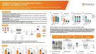 科学海报STEMprep™ Automated Tissue Dissociation System: Maximize Cell Viability and Yield
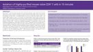 科学海报Immunomagnetic Cell Isolation of Highly Purified Mouse Naïve CD4+ T Cells in 15 Minutes

 沪公网安备31010102008431号