
 EasySep™小鼠TIL(CD45)正选试剂盒


技术资料
-
-
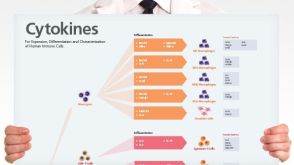 挂图Human Immune Cytokines Infographic of key cytokines for expansion, differentiation and characterization of major immune cell types
 -
-
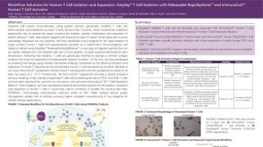
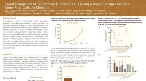 科学海报Rapid Expansion of Functional Human T Cells Using a Novel Serum-Free and Xeno-Free Culture Medium
Conference:
CCIC 2015
-
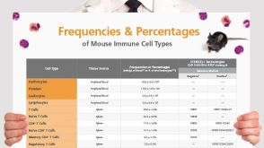 挂图Frequencies and Percentages of Mouse Immune Cell Types List of the frequencies of over 25 immune cell types in C57BL/6 mice
 -
产品号#:
18063
15361
15861
15861RF
18231
产品名:
EasySep™人CD4+CD127low CD25+调节性T细胞分选试剂盒
RosetteSep™人 CD4+ CD127low T细胞富集抗体混合物
-
-

-
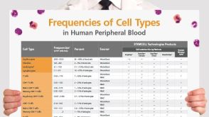 挂图血液相关来源中人细胞类型的比例 List of the frequencies of over 35 cell types in normal human blood-related sources.
 -
产品号#:
07930
07931
07940
07955
07956
07959
07954
100-1061
07952
产品名:
CryoStor® CS10
CryoStor® CS10
CryoStor® CS10
CryoStor® CS10
CryoStor® CS10
CryoStor® CS10
CryoStor® CS10

 沪公网安备31010102008431号