Berer K et al. (OCT 2017)
Proceedings of the National Academy of Sciences of the United States of America 114 40 10719--10724
Gut microbiota from multiple sclerosis patients enables spontaneous autoimmune encephalomyelitis in mice.
There is emerging evidence that the commensal microbiota has a role in the pathogenesis of multiple sclerosis (MS),a putative autoimmune disease of the CNS. Here,we compared the gut microbial composition of 34 monozygotic twin pairs discordant for MS. While there were no major differences in the overall microbial profiles,we found a significant increase in some taxa such as Akkermansia in untreated MS twins. Furthermore,most notably,when transplanted to a transgenic mouse model of spontaneous brain autoimmunity,MS twin-derived microbiota induced a significantly higher incidence of autoimmunity than the healthy twin-derived microbiota. The microbial profiles of the colonized mice showed a high intraindividual and remarkable temporal stability with several differences,including Sutterella,an organism shown to induce a protective immunoregulatory profile in vitro. Immune cells from mouse recipients of MS-twin samples produced less IL-10 than immune cells from mice colonized with healthy-twin samples. IL-10 may have a regulatory role in spontaneous CNS autoimmunity,as neutralization of the cytokine in mice colonized with healthy-twin fecal samples increased disease incidence. These findings provide evidence that MS-derived microbiota contain factors that precipitate an MS-like autoimmune disease in a transgenic mouse model. They hence encourage the detailed search for protective and pathogenic microbial components in human MS.
View Publication
产品号#:
15022
15062
产品名:
RosetteSep™人CD4+ T细胞富集抗体混合物
RosetteSep™人CD4+ T细胞富集抗体混合物
C. Gu et al. (jul 2019)
Journal of immunology (Baltimore,Md. : 1950) 203 2 389--399
Signaling Cascade through DC-ASGPR Induces Transcriptionally Active CREB for IL-10 Induction and Immune Regulation.
The types and magnitude of Ag-specific immune responses can be determined by the functional plasticity of dendritic cells (DCs). However,how DCs display functional plasticity and control host immune responses have not been fully understood. In this study,we report that ligation of DC-asialoglycoprotein receptor (DC-ASGPR),a C-type lectin receptor (CLR) expressed on human DCs,resulted in rapid activation of Syk,followed by PLCgamma2 and PKCdelta engagements. However,different from other Syk-coupled CLRs,including Dectin-1,signaling cascade through DC-ASGPR did not trigger NF-kappaB activation. Instead,it selectively activated MAPK ERK1/2 and JNK. Rapid and prolonged phosphorylation of ERK1/2 led to sequential activation of p90RSK and CREB,which consequently bound to IL10 promoter and initiated cytokine expression. In addition,DC-ASGPR ligation activated Akt,which differentially regulated the activities of GSK-3alpha/beta and beta-catenin and further contributed to IL-10 expression. Our observations demonstrate that DC-ASGPR induces IL-10 expression via an intrinsic signaling pathway,which provides a molecular explanation for DC-ASGPR-mediated programing of DCs to control host immune responses.
View Publication
S. Bhatia et al. (may 2019)
Cancer research 79 10 2722--2735
Inhibition of EphB4-Ephrin-B2 Signaling Reprograms the Tumor Immune Microenvironment in Head and Neck Cancers.
Identifying targets present in the tumor microenvironment that contribute to immune evasion has become an important area of research. In this study,we identified EphB4-ephrin-B2 signaling as a regulator of both innate and adaptive components of the immune system. EphB4 belongs to receptor tyrosine kinase family that interacts with ephrin-B2 ligand at sites of cell-cell contact,resulting in bidirectional signaling. We found that EphB4-ephrin-B2 inhibition alone or in combination with radiation (RT) reduced intratumoral regulatory T cells (Tregs) and increased activation of both CD8+ and CD4+Foxp3- T cells compared with the control group in an orthotopic head and neck squamous cell carcinoma (HNSCC) model. We also compared the effect of EphB4-ephrin-B2 inhibition combined with RT with combined anti-PDL1 and RT and observed similar tumor growth suppression,particularly at early time-points. A patient-derived xenograft model showed reduction of tumor-associated M2 macrophages and favored polarization towards an antitumoral M1 phenotype following EphB4-ephrin-B2 inhibition with RT. In vitro,EphB4 signaling inhibition decreased Ki67-expressing Tregs and Treg activation compared with the control group. Overall,our study is the first to implicate the role of EphB4-ephrin-B2 in tumor immune response. Moreover,our findings suggest that EphB4-ephrin-B2 inhibition combined with RT represents a potential alternative for patients with HNSCC and could be particularly beneficial for patients who are ineligible to receive or cannot tolerate anti-PDL1 therapy. SIGNIFICANCE: These findings present EphB4-ephrin-B2 inhibition as an alternative to anti-PDL1 therapeutics that can be used in combination with radiation to induce an effective antitumor immune response in patients with HNSCC.
View Publication
产品号#:
17952
17952RF
100-0696
产品名:
EasySep™人CD4+ T细胞分选试剂盒
RoboSep™ 人CD4+ T细胞分选试剂盒
EasySep™人CD4+ T细胞分离试剂盒
L. Hang et al. (apr 2019)
Journal of immunology (Baltimore,Md. : 1950) 202 8 2473--2481
Heligmosomoides polygyrus bakeri Infection Decreases Smad7 Expression in Intestinal CD4+ T Cells, Which Allows TGF-beta to Induce IL-10-Producing Regulatory T Cells That Block Colitis.
Helminthic infections modulate host immunity and may protect their hosts from developing immunological diseases like inflammatory bowel disease. Induction of regulatory T cells (Tregs) may be an important part of this protective process. Heligmosomoides polygyrus bakeri infection also promotes the production of the regulatory cytokines TGF-beta and IL-10 in the gut. In the intestines,TGF-beta helps induce regulatory T cells. This study used Foxp3/IL-10 double reporter mice to investigate the effect of TGF-beta on the differentiation of colon and mesenteric lymph node-derived murine Foxp3- IL-10- CD4+ T cells into their regulatory phenotypes. Foxp3- IL-10- CD4+ T cells from H. polygyrus bakeri-infected mice,as opposed to T cells from uninfected animals,cultured in vitro with TGF-beta and anti-CD3/CD28 mAb differentiated into Foxp3+ and/or IL-10+ T cells. The IL-10-producing T cells nearly all displayed CD25. Smad7 is a natural inhibitor of TGF-beta signaling. In contrast to gut T cells from uninfected mice,Foxp3- IL10- CD4+ T cells from H. polygyrus bakeri-infected mice displayed reduced Smad7 expression and responded to TGF-beta with Smad2/3 phosphorylation. The TGF-beta-induced Tregs that express IL-10 blocked colitis when transferred into the Rag/CD25- CD4+ T cell transfer model of inflammatory bowel disease. TGF-beta had a greatly diminished capacity to induce Tregs in H. polygyrus bakeri-infected transgenic mice with constitutively high T cell-specific Smad7 expression. Thus,infection with H. polygyrus bakeri causes down-modulation in Smad7 expression in intestinal CD4+ T cells,which allows the TGF-beta produced in response to the infection to induce the Tregs that prevent colitis.
View Publication
产品号#:
19751
19751RF
产品名:
B. L. Jamison et al. (jul 2019)
Journal of immunology (Baltimore,Md. : 1950) 203 1 48--57
Nanoparticles Containing an Insulin-ChgA Hybrid Peptide Protect from Transfer of Autoimmune Diabetes by Shifting the Balance between Effector T Cells and Regulatory T Cells.
CD4 T cells play a critical role in promoting the development of autoimmunity in type 1 diabetes. The diabetogenic CD4 T cell clone BDC-2.5,originally isolated from a NOD mouse,has been widely used to study the contribution of autoreactive CD4 T cells and relevant Ags to autoimmune diabetes. Recent work from our laboratory has shown that the Ag for BDC-2.5 T cells is a hybrid insulin peptide (2.5HIP) consisting of an insulin C-peptide fragment fused to a peptide from chromogranin A (ChgA) and that endogenous 2.5HIP-reactive T cells are major contributors to autoimmune pathology in NOD mice. The objective of this study was to determine if poly(lactide-co-glycolide) (PLG) nanoparticles (NPs) loaded with the 2.5HIP Ag (2.5HIP-coupled PLG NPs) can tolerize BDC-2.5 T cells. Infusion of 2.5HIP-coupled PLG NPs was found to prevent diabetes in an adoptive transfer model by impairing the ability of BDC-2.5 T cells to produce proinflammatory cytokines through induction of anergy,leading to an increase in the ratio of Foxp3+ regulatory T cells to IFN-gamma+ effector T cells. To our knowledge,this work is the first to use a hybrid insulin peptide,or any neoepitope,to re-educate diabetogenic T cells and may have significant implications for the development of an Ag-specific therapy for type 1 diabetes patients.
View Publication
产品号#:
19852
19852RF
18783
18783RF
18765
18765RF
产品名:
EasySep™小鼠CD4+ T细胞分选试剂盒
RoboSep™ 小鼠CD4+ T细胞分选试剂盒
EasySep™小鼠CD4+CD25+调节性T细胞分选试剂盒II
RoboSep™ 小鼠CD4+CD25+调节性T细胞分选试剂盒II
EasySep™小鼠CD4+ CD62L+ T细胞分选试剂盒
RoboSep™ 小鼠CD4+ CD62L+ T细胞分选试剂盒
S. Omenetti et al. (jun 2019)
Immunity
The Intestine Harbors Functionally Distinct Homeostatic Tissue-Resident and Inflammatory Th17 Cells.
T helper 17 (Th17) cells are pathogenic in many inflammatory diseases,but also support the integrity of the intestinal barrier in a non-inflammatory manner. It is unclear what distinguishes inflammatory Th17 cells elicited by pathogens and tissue-resident homeostatic Th17 cells elicited by commensals. Here,we compared the characteristics of Th17 cells differentiating in response to commensal bacteria (SFB) to those differentiating in response to a pathogen (Citrobacter rodentium). Homeostatic Th17 cells exhibited little plasticity towards expression of inflammatory cytokines,were characterized by a metabolism typical of quiescent or memory T cells,and did not participate in inflammatory processes. In contrast,infection-induced Th17 cells showed extensive plasticity towards pro-inflammatory cytokines,disseminated widely into the periphery,and engaged aerobic glycolysis in addition to oxidative phosphorylation typical for inflammatory effector cells. These findings will help ensure that future therapies directed against inflammatory Th17 cells do not inadvertently damage the resident gut population.
View Publication
Friesen TJ et al. (MAY 2016)
The Journal of Experimental Medicine 213 6 913--920
Recent thymic emigrants are tolerized in the absence of inflammation.
T cell development requires a period of postthymic maturation. Why this is the case has remained a mystery,particularly given the rigors of intrathymic developmental checkpoints,successfully traversed by only ∼5% of thymocytes. We now show that the first few weeks of T cell residence in the lymphoid periphery define a period of heightened susceptibility to tolerance induction to tissue-restricted antigens (TRAs),the outcome of which depends on the context in which recent thymic emigrants (RTEs) encounter antigen. After encounter with TRAs in the absence of inflammation,RTEs exhibited defects in proliferation,diminished cytokine production,elevated expression of anergy-associated genes,and diminished diabetogenicity. These properties were mirrored in vitro by enhanced RTE susceptibility to regulatory T cell-mediated suppression. In the presence of inflammation,RTEs and mature T cells were,in contrast,equally capable of inducing diabetes,proliferating,and producing cytokines. Thus,recirculating RTEs encounter TRAs during a transitional developmental stage that facilitates tolerance induction,but inflammation converts antigen-exposed,tolerance-prone RTEs into competent effector cells.
View Publication
产品号#:
19852
19852RF
19853
19853RF
产品名:
EasySep™小鼠CD4+ T细胞分选试剂盒
RoboSep™ 小鼠CD4+ T细胞分选试剂盒
EasySep™小鼠CD8+ T细胞分选试剂盒
RoboSep™ 小鼠CD8+ T细胞分选试剂盒
Apps R et al. (MAY 2016)
Cell Host & Microbe 19 5 686--95
HIV-1 Vpu Mediates HLA-C Downregulation.
Many pathogens evade cytotoxic T lymphocytes (CTLs) by downregulating HLA molecules on infected cells,but the loss of HLA can trigger NK cell-mediated lysis. HIV-1 is thought to subvert CTLs while preserving NK cell inhibition by Nef-mediated downregulation of HLA-A and -B but not HLA-C molecules. We find that HLA-C is downregulated by most primary HIV-1 clones,including transmitted founder viruses,in contrast to the laboratory-adapted NL4-3 virus. HLA-C reduction is mediated by viral Vpu and reduces the ability of HLA-C restricted CTLs to suppress viral replication in CD4+ cells in vitro. HLA-A/B are unaffected by Vpu,and primary HIV-1 clones vary in their ability to downregulate HLA-C,possibly in response to whether CTLs or NK cells dominate immune pressure through HLA-C. HIV-2 also suppresses HLA-C expression through distinct mechanisms,underscoring the immune pressure HLA-C exerts on HIV. This viral immune evasion casts new light on the roles of CTLs and NK cells in immune responses against HIV.
View Publication
产品号#:
17952
17952RF
100-0696
产品名:
EasySep™人CD4+ T细胞分选试剂盒
RoboSep™ 人CD4+ T细胞分选试剂盒
EasySep™人CD4+ T细胞分离试剂盒
Jung Y et al. (SEP 2016)
Proceedings of the National Academy of Sciences of the United States of America
Three-dimensional localization of T-cell receptors in relation to microvilli using a combination of superresolution microscopies.
Leukocyte microvilli are flexible projections enriched with adhesion molecules. The role of these cellular projections in the ability of T cells to probe antigen-presenting cells has been elusive. In this study,we probe the spatial relation of microvilli and T-cell receptors (TCRs),the major molecules responsible for antigen recognition on the T-cell membrane. To this end,an effective and robust methodology for mapping membrane protein distribution in relation to the 3D surface structure of cells is introduced,based on two complementary superresolution microscopies. Strikingly,TCRs are found to be highly localized on microvilli,in both peripheral blood human T cells and differentiated effector T cells,and are barely found on the cell body. This is a decisive demonstration that different types of T cells universally localize their TCRs to microvilli,immediately pointing to these surface projections as effective sensors for antigenic moieties. This finding also suggests how previously reported membrane clusters might form,with microvilli serving as anchors for specific T-cell surface molecules.
View Publication


 EasySep™小鼠TIL(CD45)正选试剂盒





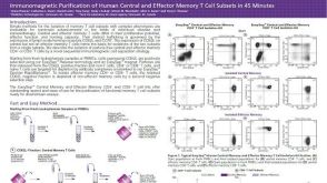 科学海报Immunomagnetic Purification of Human Central and Effector Memory T Cell Subsets in 45 Minutes
 沪公网安备31010102008431号