Discrimination of polycythemias and thrombocytoses by novel, simple, accurate clonality assays and comparison with PRV-1 expression and BFU-E response to erythropoietin.
Essential thrombocythemia (ET) and polycythemia vera (PV) are clonal myeloproliferative disorders that are often difficult to distinguish from other causes of elevated blood cell counts. Assays that could reliably detect clonal hematopoiesis would therefore be extremely valuable for diagnosis. We previously reported 3 X-chromosome transcription-based clonality assays (TCAs) involving the G6PD,IDS,and MPP1 genes,which together were informative in about 65% of female subjects. To increase our ability to detect clonality,we developed simple TCA for detecting the transcripts of 2 additional X-chromosome genes: Bruton tyrosine kinase (BTK) and 4-and-a-half LIM domain 1 (FHL1). The combination of TCA established the presence or absence of clonal hematopoiesis in about 90% of female subjects. We show that both genes are subject to X-chromosome inactivation and are polymorphic in all major US ethnic groups. The 5 TCAs were used to examine clonality in 46 female patients along with assays for erythropoietin-independent erythroid colonies (EECs) and granulocyte PRV-1 mRNA levels to discriminate polycythemias and thrombocytoses. Of these,all 19 patients with familial polycythemia or thrombocytosis had polyclonal hematopoiesis,whereas 22 of 26 patients with clinical evidence of myeloproliferative disorder and 1 patient with clinically obscure polycythemia were clonal. Interestingly,interferon alpha therapy in 2 patients with PV was associated with reversion of clonal to polyclonal hematopoiesis. EECs were observed in 14 of 14 patients with PV and 4 of 12 with ET,and increased granulocyte PRV-1 mRNA levels were found in 9 of 13 patients with PV and 2 of 12 with ET. Thus,these novel clonality assays are useful in the diagnosis and follow-up of polycythemic conditions and disorders with increased platelet levels.
View Publication
产品号#:
04531
15021
15061
产品名:
MethoCult™ H4531
RosetteSep™人T细胞富集抗体混合物
RosetteSep™人T细胞富集抗体混合物
Deonarain R et al. (NOV 2003)
Proceedings of the National Academy of Sciences of the United States of America 100 23 13453--8
Critical roles for IFN-beta in lymphoid development, myelopoiesis, and tumor development: links to tumor necrosis factor alpha.
We have generated mice null for IFN-beta and report the diverse consequences of IFN-beta for both the innate and adaptive arms of immunity. Despite no abnormalities in the proportional balance of CD4 and CD8 T cell populations in the peripheral blood,thymus,and spleen of IFN-beta-/- mice,activated lymph node and splenic T lymphocytes exhibit enhanced T cell proliferation and decreased tumor necrosis factor alpha production,relative to IFN-beta+/+ mice. Notably,constitutive and induced expression of tumor necrosis factor alpha is reduced in the spleen and bone marrow (BM) macrophages,respectively,of IFN-beta-/- mice. We also observe an altered splenic architecture in IFN-beta-/- mice and a reduction in resident macrophages. We identify a potential defect in B cell maturation in IFN-beta-/- mice,associated with a decrease in B220+ve/high/CD43-ve BM-derived cells and a reduction in BP-1,IgM,and CD23 expression. Circulating IgM-,Mac-1-,and Gr-1-positive cells are also substantially decreased in IFN-beta-/- mice. The decrease in the numbers of circulating macrophages and granulocytes likely reflects defective maturation of primitive BM hematopoiesis in mice,shown by the reduction of colony-forming units,granulocyte-macrophage. We proceeded to evaluate the in vivo growth of malignant cells in the IFN-beta-/- background and give evidence that Lewis lung carcinoma-specific tumor growth is more aggressive in IFN-beta-/- mice. Taken altogether,our data suggest that,in addition to the direct growth-inhibitory effects on tumor cells,IFN-beta is required during different stages of maturation in the development of the immune system.
View Publication
产品号#:
03434
03444
产品名:
MethoCult™ GF M3434
MethoCult™ GF M3434
Storck S et al. (FEB 2005)
Molecular and cellular biology 25 4 1437--45
Normal immune system development in mice lacking the Deltex-1 RING finger domain.
The Notch signaling pathway controls several cell fate decisions during lymphocyte development,from T-cell lineage commitment to the peripheral differentiation of B and T lymphocytes. Deltex-1 is a RING finger ubiquitin ligase which is conserved from Drosophila to humans and has been proposed to be a regulator of Notch signaling. Its pattern of lymphoid expression as well as gain-of-function experiments suggest that Deltex-1 regulates both B-cell lineage and splenic marginal-zone B-cell commitment. Deltex-1 was also found to be highly expressed in germinal-center B cells. To investigate the physiological function of Deltex-1,we generated a mouse strain lacking the Deltex-1 RING finger domain,which is essential for its ubiquitin ligase activity. Deltex-1(Delta/Delta) mice were viable and fertile. A detailed histological analysis did not reveal any defects in major organs. T- and B-cell development was normal,as were humoral responses against T-dependent and T-independent antigens. These data indicate that the Deltex-1 ubiquitin ligase activity is dispensable for mouse development and immune function. Possible compensatory mechanisms,in particular those from a fourth Deltex gene identified during the course of this study,are also discussed.
View Publication
产品号#:
18751
18751RF
产品名:
Vieillard V et al. (AUG 2005)
Proceedings of the National Academy of Sciences 102 31 10981--86
NK cytotoxicity against CD4+ T cells during HIV-1 infection: A gp41 peptide induces the expression of an NKp44 ligand
HIV infection leads to a state of chronic immune activation and progressive deterioration in immune function,manifested most recognizably by the progressive depletion of CD4+ T cells. A substantial percentage of natural killer (NK) cells from patients with HIV infection are activated and express the natural cytotoxicity receptor (NCR) NKp44. Here we show that a cellular ligand for NKp44 (NKp44L) is expressed during HIV-1 infection and is correlated with both the progression of CD4+ T cell depletion and the increase of viral load. CD4+ T cells expressing this ligand are highly sensitive to the NK lysis activity mediated by NKp44+ NK cells. The expression of NKp44L is induced by the linear motif NH2-SWSNKS-COOH of the HIV-1 envelope gp41 protein. This highly conserved motif appears critical to the sharp increase in NK lysis of CD4+ T cells from HIV-infected patients. These studies strongly suggest that induction of NKp44L plays a key role in the lysis of CD4+ T cells by activated NK cells in HIV infection and consequently provide a framework for considering how HIV-1 may use NK cell immune surveillance to trigger CD4+ T cells. Understanding this mechanism may help to develop future therapeutic strategies and vaccines against HIV-1 infection.
View Publication
产品号#:
03800
03801
03802
03803
03804
03805
03806
05150
15021
15061
产品名:
ClonaCell™-HY杂交瘤试剂盒
ClonaCell™-HY培养基A
ClonaCell™-HY 培养基 B
ClonaCell™-HY 培养基 C
ClonaCell™-HY 培养基 D
ClonaCell™-HY 培养基 E
ClonaCell™-HY PEG
MyeloCult™ H5100
RosetteSep™人T细胞富集抗体混合物
RosetteSep™人T细胞富集抗体混合物
Li Q et al. (AUG 2005)
Proceedings of the National Academy of Sciences of the United States of America 102 35 12425--30
Enhanced NF-kappaB activation and cellular function in macrophages lacking IkappaB kinase 1 (IKK1).
IkappaB kinase (IKK) complex plays a key regulatory role in macrophages for NF-kappaB activation during both innate and adaptive immune responses. Because IKK1-/- mice died at birth,we differentiated functional macrophages from embryonic day 15.5 IKK1 mutant embryonic liver. The embryonic liver-derived macrophage (ELDM) showed enhanced phagocytotic clearance of bacteria,more efficient antigen-presenting capacity,elevated secretion of several key proinflammatory cytokines and chemokines,and known NFkappaB target genes. Increased NFkappaB activity in IKK1 mutant ELDM was the result of prolonged degradation of IkappaBalpha in response to infectious pathogens. The delayed restoration of IkappaBalpha in pathogen-activated IKK1-/- ELDM was a direct consequence of uncontrolled IKK2 kinase activity. We hypothesize that IKK1 plays a checkpoint role in the proper control of IkappaBalpha kinase activity in innate and adaptive immunity.
View Publication
产品号#:
02532
02832
09600
09650
产品名:
StemSpan™ SFEM
StemSpan™ SFEM
Maes C et al. (MAY 2006)
The Journal of clinical investigation 116 5 1230--42
Placental growth factor mediates mesenchymal cell development, cartilage turnover, and bone remodeling during fracture repair.
Current therapies for delayed- or nonunion bone fractures are still largely ineffective. Previous studies indicated that the VEGF homolog placental growth factor (PlGF) has a more significant role in disease than in health. Therefore we investigated the role of PlGF in a model of semi-stabilized bone fracture healing. Fracture repair in mice lacking PlGF was impaired and characterized by a massive accumulation of cartilage in the callus,reminiscent of delayed- or nonunion fractures. PlGF was required for the early recruitment of inflammatory cells and the vascularization of the fracture wound. Interestingly,however,PlGF also played a role in the subsequent stages of the repair process. Indeed in vivo and in vitro findings indicated that PlGF induced the proliferation and osteogenic differentiation of mesenchymal progenitors and stimulated cartilage turnover by particular MMPs. Later in the process,PlGF was required for the remodeling of the newly formed bone by stimulating osteoclast differentiation. As PlGF expression was increased throughout the process of bone repair and all the important cell types involved expressed its receptor VEGFR-1,the present data suggest that PlGF is required for mediating and coordinating the key aspects of fracture repair. Therefore PlGF may potentially offer therapeutic advantages for fracture repair.
View Publication


 EasySep™小鼠TIL(CD45)正选试剂盒




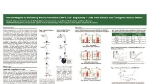 科学海报Cell Isolation of Functional CD4+CD25+ Regulatory T Cells from Mouse Strains
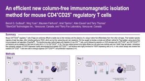 科学海报Immunomagnetic Isolation Method for Mouse CD4+CD25+ Regulatory T Cells
 科学海报One-Step Enrichment of Leukocyte Subsets Directly in the Blood Collection Tube


 沪公网安备31010102008431号