Kabanova A et al. (APR 2016)
Cell Reports 15 1 9--18
Human Cytotoxic T Lymphocytes Form Dysfunctional Immune Synapses with B Cells Characterized by Non-Polarized Lytic Granule Release.
Suppression of the cytotoxic T cell (CTL) immune response has been proposed as one mechanism for immune evasion in cancer. In this study,we have explored the underlying basis for CTL suppression in the context of B cell malignancies. We document that human B cells have an intrinsic ability to resist killing by freshly isolated cytotoxic T cells (CTLs),but are susceptible to lysis by IL-2 activated CTL blasts and CTLs isolated from immunotherapy-treated patients with chronic lymphocytic leukemia (CLL). Impaired killing was associated with the formation of dysfunctional non-lytic immune synapses characterized by the presence of defective linker for activation of T cells (LAT) signaling and non-polarized release of the lytic granules transported by ADP-ribosylation factor-like protein 8 (Arl8). We propose that non-lytic degranulation of CTLs are a key regulatory mechanism of evasion through which B cells may interfere with the formation of functional immune synapses by CTLs.
View Publication
Webb CF et al. (MAR 2011)
Molecular and cellular biology 31 5 1041--53
The ARID family transcription factor bright is required for both hematopoietic stem cell and B lineage development.
Bright/Arid3a has been characterized both as an activator of immunoglobulin heavy-chain transcription and as a proto-oncogene. Although Bright expression is highly B lineage stage restricted in adult mice,its expression in the earliest identifiable hematopoietic stem cell (HSC) population suggests that Bright might have additional functions. We showed that textgreater99% of Bright(-/-) embryos die at midgestation from failed hematopoiesis. Bright(-/-) embryonic day 12.5 (E12.5) fetal livers showed an increase in the expression of immature markers. Colony-forming assays indicated that the hematopoietic potential of Bright(-/-) mice is markedly reduced. Rare survivors of lethality,which were not compensated by the closely related paralogue Bright-derived protein (Bdp)/Arid3b,suffered HSC deficits in their bone marrow as well as B lineage-intrinsic developmental and functional deficiencies in their peripheries. These include a reduction in a natural antibody,B-1 responses to phosphocholine,and selective T-dependent impairment of IgG1 class switching. Our results place Bright/Arid3a on a select list of transcriptional regulators required to program both HSC and lineage-specific differentiation.
View Publication
Activation-induced cytidine deaminase deficiency accelerates autoimmune diabetes in NOD mice.
B cells play an important role in type 1 diabetes (T1D) development. However,the role of B cell activation-induced cytidine deaminase (AID) in diabetes development is not clear. We hypothesized that AID is important in the immunopathogenesis of T1D. To test this hypothesis,we generated AID-deficient (AID-/-) NOD mice. We found that AID-/-NOD mice developed accelerated T1D,with worse insulitis and high levels of anti-insulin autoantibody in the circulation. Interestingly,neither maternal IgG transferred through placenta,nor IgA transferred through milk affected the accelerated diabetes development. AID-/-NOD mice showed increased activation and proliferation of B and T cells. We found enhanced T-B cell interactions in AID-/-NOD mice,with increased T-bet and IFN-γ expression in CD4+ T cells in the presence of AID-/- B cells. Moreover,excessive lymphoid expansion was observed in AID-/-NOD mice. Importantly,antigen-specific BDC2.5 CD4+ T cells caused more rapid onset of diabetes when cotransferred with AID-/- B cells than when cotransferred with AID+/+ B cells. Thus,our study provides insights into the role of AID in T1D. Our data also suggest that AID is a negative regulator of immune tolerance and ablation of AID can lead to exacerbated islet autoimmunity and accelerated T1D development.
View Publication
产品号#:
19854
19854RF
产品名:
EasySep™小鼠B细胞分选试剂盒
RoboSep™ 小鼠B细胞分选试剂盒
挂图
Human Immune Cytokines
Infographic of key cytokines for expansion, differentiation and characterization of major immune cell types

 EasySep™小鼠TIL(CD45)正选试剂盒


 科学海报Immunomagnetic Cell Isolation of Untouched Lymphocyte Populations Directly from Whole Blood
 实验方案Optimizing Delivery Efficiency with Fluorescent Dextran Using the CellPore™ Transfection System

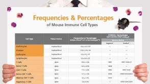 挂图Frequencies and Percentages of Mouse Immune Cell Types List of the frequencies of over 25 immune cell types in C57BL/6 mice
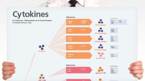 挂图Human Immune Cytokines Infographic of key cytokines for expansion, differentiation and characterization of major immune cell types

 沪公网安备31010102008431号