Allan LL et al. (MAY 2011)
Journal of immunology (Baltimore,Md. : 1950) 186 9 5261--72
CD1d and CD1c expression in human B cells is regulated by activation and retinoic acid receptor signaling.
B cell activation and Ab production in response to protein Ags requires presentation of peptides for recruitment of T cell help. We and others have recently demonstrated that B cells can also acquire innate help by presenting lipid Ags via CD1d to NKT cells. Given the newfound contribution of NKT cells to humoral immunity,we sought to identify the pathways that regulate CD1 molecule expression in human B cells. We show that ex vivo,activated and memory B cells expressed lower levels of CD1d compared with resting,naive,and marginal zone-like B cells. In vitro,CD1d was downregulated by all forms of B cell activation,leaving a narrow temporal window in which B cells could activate NKT cells. CD1c expression and function also decreased following activation by CD40L alone,whereas activation via the BCR significantly upregulated CD1c,particularly on marginal zone-like B cells. We found that the CD40L-induced downregulation of CD1d and CD1c correlated with diminished expression of retinoic acid receptor α (RARα) response genes,an effect that was reversed by RARα agonists. However,BCR-induced upregulation of CD1c was independent of the RAR pathway. Our findings that both CD1d and CD1c are upregulated by RARα signaling in human B cells is distinct from effects reported in dendritic cells,in which CD1c is inversely downregulated. One functional consequence of CD1d upregulation by retinoic acid was NKT cell cytotoxicity toward B cells. These results are central to our understanding of how CD1-restricted T cells may control humoral immunity.
View Publication
产品号#:
01700
01705
18054
18054RF
01702
产品名:
ALDEFLUOR™ 试剂盒
ALDEFLUOR™ DEAB试剂, 1.5 mM, 1 mL
ALDEFLUOR™检测缓冲液
Le Y et al. (MAR 2005)
Journal of immunology (Baltimore,Md. : 1950) 174 5 2582--90
CXC chemokine ligand 12-induced focal adhesion kinase activation and segregation into membrane domains is modulated by regulator of G protein signaling 1 in pro-B cells.
CXCL12-induced chemotaxis and adhesion to VCAM-1 decrease as B cells differentiate in the bone marrow. However,the mechanisms that regulate CXCL12/CXCR4-mediated signaling are poorly understood. We report that after CXCL12 stimulation of progenitor B cells,focal adhesion kinase (FAK) and PI3K are inducibly recruited to raft-associated membrane domains. After CXCL12 stimulation,phosphorylated FAK is also localized in membrane domains. The CXCL12/CXCR4-FAK pathway is membrane cholesterol dependent and impaired by metabolic inhibitors of G(i),Src family,and the GTPase-activating protein,regulator of G protein signaling 1 (RGS1). In the bone marrow,RGS1 mRNA expression is low in progenitor B cells and high in mature B cells,implying developmental regulation of CXCL12/CXCR4 signaling by RGS1. CXCL12-induced chemotaxis and adhesion are impaired when FAK recruitment and phosphorylation are inhibited by either membrane cholesterol depletion or overexpression of RGS1 in progenitor B cells. We conclude that the recruitment of signaling molecules to specific membrane domains plays an important role in CXCL12/CXCR4-induced cellular responses.
View Publication
产品号#:
产品名:
Balakrishnan K et al. (OCT 2006)
Blood 108 7 2392--8
Forodesine, an inhibitor of purine nucleoside phosphorylase, induces apoptosis in chronic lymphocytic leukemia cells.
Purine nucleoside phosphorylase (PNP) deficiency in humans results in T lymphocytopenia. Forodesine,a potent inhibitor of PNP,was designed based on the transition-state structure stabilized by the enzyme. Previous studies established that forodesine in the presence of deoxyguanosine (dGuo) inhibits the proliferation of T lymphocytes. A phase 1 clinical trial of forodesine in T-cell malignancies demonstrated significant antileukemic activity with an increase in intracellular dGuo triphosphate (dGTP). High accumulation of dGTP in T cells may be dependent on the levels of deoxynucleoside kinases. Because B-cell chronic lymphocytic leukemia (B-CLL) cells have high activity of deoxycytidine kinase (dCK),we hypothesized that these lymphocytes would respond to forodesine. This postulate was tested in primary lymphocytes during in vitro investigations. Lymphocytes from 12 patients with CLL were incubated with forodesine and dGuo. These CLL cells showed a wide variation in the accumulation of intracellular dGTP without any effect on other deoxynucleotides. This was associated with DNA damage-induced p53 stabilization,phosphorylation of p53 at Ser15,and activation of p21. The dGTP accumulation was related to induction of apoptosis measured by caspase activation,changes in mitochondrial membrane potential,and PARP cleavage. Based on these data,a phase 2 clinical trial of forodesine has been initiated for CLL patients.
View Publication
产品号#:
19051
19051RF
19054
19054RF
产品名:
EasySep™人T细胞富集试剂盒
RoboSep™ 人T细胞富集试剂盒含滤芯吸头
EasySep™人B细胞富集试剂盒
RoboSep™ 人B细胞富集试剂盒含滤芯吸头
Finstad SL et al. (JUL 2007)
Journal of virology 81 13 7274--9
Diminished potential for B-lymphoid differentiation after murine leukemia virus infection in vivo and in EML hematopoietic progenitor cells.
Infection with a recombinant murine-feline gammaretrovirus,MoFe2,or with the parent virus,Moloney murine leukemia virus,caused significant reduction in B-lymphoid differentiation of bone marrow at 2 to 8 weeks postinfection. The suppression was selective,in that myeloid potential was significantly increased by infection. Analysis of cell surface markers and immunoglobulin H gene rearrangements in an in vitro model demonstrated normal B-lymphoid differentiation after infection but significantly reduced viability of differentiating cells. This reduction in viability may confer a selective advantage on undifferentiated lymphoid progenitors in the bone marrow of gammaretrovirus-infected animals and thereby contribute to the establishment of a premalignant state.
View Publication
产品号#:
03630
03434
03444
产品名:
MethoCult™ M3630
MethoCult™ GF M3434
MethoCult™ GF M3434
Nguyen CQ et al. (JUL 2007)
Journal of immunology (Baltimore,Md. : 1950) 179 1 382--90
IL-4-STAT6 signal transduction-dependent induction of the clinical phase of Sjögren's syndrome-like disease of the nonobese diabetic mouse.
NOD.B10-H2(b) and NOD/LtJ mice manifest,respectively,many features of primary and secondary Sjögren's syndrome (SjS),an autoimmune disease affecting primarily the salivary and lacrimal glands leading to xerostomia (dry mouth) and xerophthalmia (dry eyes). B lymphocytes play a central role in the onset of SjS with clinical manifestations dependent on the appearance of autoantibodies reactive to multiple components of acinar cells. Previous studies with NOD.IL4(-/-) and NOD.B10-H2(b).IL4(-/-) mice suggest that the Th2 cytokine,IL-4,plays a vital role in the development and onset of SjS-like disease in the NOD mouse model. To investigate the molecular mechanisms by which IL-4 controls SjS development,a Stat6 gene knockout mouse,NOD.B10-H2(b).C-Stat6(-/-),was constructed and its disease profile was defined and compared with that of NOD.B10-H2(b).C-Stat6(+/+) mice. As the NOD.B10-H2(b).C-Stat6(-/-) mice aged from 4 to 24 wk,they exhibited leukocyte infiltration of the exocrine glands,produced anti-nuclear autoantibodies,and showed loss and gain of saliva-associated proteolytic enzymes,similar to NOD.B10-H2(b).C-Stat6(+/+) mice. In contrast,NOD.B10-H2(b).C-Stat6(-/-) mice failed to develop glandular dysfunction,maintaining normal saliva flow rates. NOD.B10-H2(b).C-Stat6(-/-) mice were found to lack IgG1 isotype-specific anti-muscarinic acetylcholine type-3 receptor autoantibodies. Furthermore,the IgG fractions from NOD.B10-H2(b).C-Stat6(-/-) sera were unable to induce glandular dysfunction when injected into naive recipient C57BL/6 mice. NOD.B10-H2(b).C-Stat6(-/-) mice,like NOD.B10-H2(b).IL4(-/-) mice,are unable to synthesize IgG1 Abs,an observation that correlates with an inability to develop end-stage clinical SjS-like disease. These data imply a requirement for the IL-4/STAT6-pathway for onset of the clinical phase of SjS-like disease in the NOD mouse model.
View Publication

 EasySep™小鼠TIL(CD45)正选试剂盒


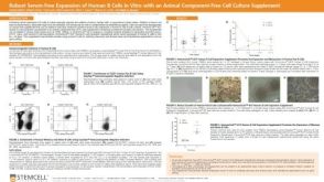 科学海报Robust Serum-Free Expansion of Human B Cells In Vitro with an Animal Component-Free Cell Culture Supplement
 技术公告Achieve Scalable, High-Quality Nucleic Acid Extractions with the EasySep™ Total Nucleic Acid Extraction Kit


 沪公网安备31010102008431号