Chou S-J et al. (APR 2017)
International journal of cardiology 232 255--263
Energy utilization of induced pluripotent stem cell-derived cardiomyocyte in Fabry disease.
BACKGROUND Fabry disease (FD) is a lysosomal storage disease in which glycosphingolipids (GB3) accumulate in organs of the human body,leading to idiopathic hypertrophic cardiomyopathy and target organ damage. Its pathophysiology is still poorly understood. OBJECTIVES We aimed to generate patient-specific induced pluripotent stem cells (iPSC) from FD patients presenting cardiomyopathy to determine whether the model could recapitulate key features of the disease phenotype and to investigate the energy metabolism in Fabry disease. METHODS Peripheral blood mononuclear cells from a 30-year-old Chinese man with a diagnosis of Fabry disease,GLA gene (IVS4+919G>A) mutation were reprogrammed into iPSCs and differentiated into iPSC-CMs and energy metabolism was analyzed in iPSC-CMs. RESULTS The FD-iPSC-CMs recapitulated numerous aspects of the FD phenotype including reduced GLA activity,cellular hypertrophy,GB3 accumulation and impaired contractility. Decreased energy metabolism with energy utilization shift to glycolysis was observed,but the decreased energy metabolism was not modified by enzyme rescue replacement (ERT) in FD-iPSCs-CMs. CONCLUSION This model provided a promising in vitro model for the investigation of the underlying disease mechanism and development of novel therapeutic strategies for FD. This potential remedy for enhancing the energetic network and utility efficiency warrants further study to identify novel therapies for the disease.
View Publication
Mariotti J et al. (JAN 2008)
Journal of immunology (Baltimore,Md. : 1950) 180 1 89--105
Ex vivo rapamycin generates apoptosis-resistant donor Th2 cells that persist in vivo and prevent hemopoietic stem cell graft rejection.
Because ex vivo rapamycin generates murine Th2 cells that prevent Graft-versus-host disease more potently than control Th2 cells,we hypothesized that rapamycin would generate Th2/Tc2 cells (Th2/Tc2.R cells) that abrogate fully MHC-disparate hemopoietic stem cell rejection more effectively than control Th2/Tc2 cells. In a B6-into-BALB/c graft rejection model,donor Th2/Tc2.R cells were indeed enriched in their capacity to prevent rejection; importantly,highly purified CD4+ Th2.R cells were also highly efficacious for preventing rejection. Rapamycin-generated Th2/Tc2 cells were less likely to die after adoptive transfer,accumulated in vivo at advanced proliferative cycles,and were present in 10-fold higher numbers than control Th2/Tc2 cells. Th2.R cells had a multifaceted,apoptosis-resistant phenotype,including: 1) reduced apoptosis after staurosporine addition,serum starvation,or CD3/CD28 costimulation; 2) reduced activation of caspases 3 and 9; and 3) increased anti-apoptotic Bcl-xL expression and reduced proapoptotic Bim and Bid expression. Using host-versus-graft reactivity as an immune correlate of graft rejection,we found that the in vivo efficacy of Th2/Tc2.R cells 1) did not require Th2/Tc2.R cell expression of IL-4,IL-10,perforin,or Fas ligand; 2) could not be reversed by IL-2,IL-7,or IL-15 posttransplant therapy; and 3) was intact after therapy with Th2.R cells relatively devoid of Foxp3 expression. We conclude that ex vivo rapamycin generates Th2 cells that are resistant to apoptosis,persist in vivo,and effectively prevent rejection by a mechanism that may be distinct from previously described graft-facilitating T cells.
View Publication
产品号#:
09600
09650
产品名:
StemSpan™ SFEM
StemSpan™ SFEM
Crist SA et al. (APR 2008)
Blood 111 7 3553--61
Nuclear factor of activated T cells (NFAT) mediates CD154 expression in megakaryocytes.
Platelets are an abundant source of CD40 ligand (CD154),an immunomodulatory and proinflammatory molecule implicated in the onset and progression of several inflammatory diseases,including systemic lupus erythematosus (SLE),diabetes,and cardiovascular disease. Heretofore considered largely restricted to activated T cells,we initiated studies to investigate the source and regulation of platelet-associated CD154. We found that CD154 is abundantly expressed in platelet precursor cells,megakaryocytes. We show that CD154 is expressed in primary human CD34+ and murine hematopoietic precursor cells only after cytokine-driven megakaryocyte differentiation. Furthermore,using several established megakaryocyte-like cells lines,we performed promoter analysis of the CD154 gene and found that NFAT,a calcium-dependent transcriptional regulator associated with activated T cells,mediated both differentiation-dependent and inducible megakaryocyte-specific CD154 expression. Overall,these data represent the first investigation of the regulation of a novel source of CD154 and suggests that platelet-associated CD154 can be biochemically modulated.
View Publication
产品号#:
09600
09650
产品名:
StemSpan™ SFEM
StemSpan™ SFEM
Sessarego N et al. (MAR 2008)
Haematologica 93 3 339--46
Multipotent mesenchymal stromal cells from amniotic fluid: solid perspectives for clinical application.
BACKGROUND: Mesenchymal stromal cells are multipotent cells considered to be of great promise for use in regenerative medicine. However,the cell dose may be a critical factor in many clinical conditions and the yield resulting from the ex vivo expansion of mesenchymal stromal cells derived from bone marrow may be insufficient. Thus,alternative sources of mesenchymal stromal cells need to be explored. In this study,mesenchymal stromal cells were successfully isolated from second trimester amniotic fluid and analyzed for chromosomal stability to validate their safety for potential utilization as a cell therapy product. DESIGN AND METHODS: Mesenchymal stromal cells were expanded up to the sixth passage starting from amniotic fluid using different culture conditions to optimize large-scale production. RESULTS: The highest number of mesenchymal stromal cells derived from amniotic fluid was reached at a low plating density; in these conditions the expansion of mesenchymal stromal cells from amniotic fluid was significantly greater than that of adult bone marrow-derived mesenchymal stromal cells. Mesenchymal stromal cells from amniotic fluid represent a relatively homogeneous population of immature cells with immunosuppressive properties and extensive proliferative potential. Despite their high proliferative capacity in culture,we did not observe any karyotypic abnormalities or transformation potential in vitro nor any tumorigenic effect in vivo. CONCLUSIONS: Fetal mesenchymal stromal cells can be extensively expanded from amniotic fluid,showing no karyotypic abnormalities or transformation potential in vitro and no tumorigenic effect in vivo. They represent a relatively homogeneous population of immature mesenchymal stromal cells with long telomeres,immunosuppressive properties and extensive proliferative potential. Our results indicate that amniotic fluid represents a rich source of mesenchymal stromal cells suitable for banking to be used when large amounts of cells are required.
View Publication
Alkaline phosphatase-positive colony formation is a sensitive, specific, and quantitative indicator of undifferentiated human embryonic stem cells.
Human embryonic stem cells (hESCs) can be maintained in vitro as immortal pluripotent cells but remain responsive to many differentiation-inducing signals. Investigation of the initial critical events involved in differentiation induction would be greatly facilitated if a specific,robust,and quantitative assay for pluripotent hESCs with self-renewal potential were available. Here we describe the results of a series of experiments to determine whether the formation of adherent alkaline phosphatase-positive (AP(+)) colonies under conditions optimized for propagating undifferentiated hESCs would meet this need. The findings can be summarized as follows. (a) Most colonies obtained under these conditions consist of textgreateror=30 AP(+) cells that coexpress OCT4,NANOG,SSEA3,SSEA4,TRA-1-60,and TRA-1-81. (b) Most such colonies are derived from SSEA3(+) cells. (c) Primary colonies contain cells that produce secondary colonies of the same composition,including cells that initiate multilineage differentiation in embryoid bodies (EBs). (d) Colony formation is independent of plating density or the colony-forming cell (CFC) content of the test population over a wide range of cell concentrations. (e) CFC frequencies decrease when differentiation is induced by exposure either to retinoic acid or to conditions that stimulate EB formation. Interestingly,this loss of AP(+) clonogenic potential also occurs more rapidly than the loss of SSEA3 or OCT4 expression. The CFC assay thus provides a simple,reliable,broadly applicable,and highly specific functional assay for quantifying undifferentiated hESCs with self-renewal potential. Its use under standardized assay conditions should enhance future elucidation of the mechanisms that regulate hESC propagation and their early differentiation.
View Publication
产品号#:
05850
05857
05870
05875
07923
36254
85850
85857
85870
85875
产品名:
Dispase (1 U/mL)
DMEM/F-12 with 15 mM HEPES
mTeSR™1
mTeSR™1
Schü et al. (MAY 2008)
Blood 111 9 4532--41
The MADS transcription factor Mef2c is a pivotal modulator of myeloid cell fate.
Mef2c is a MADS (MCM1-agamous-deficient serum response factor) transcription factor best known for its role in muscle and cardiovascular development. A causal role of up-regulated MEF2C expression in myelomonocytic acute myeloid leukemia (AML) has recently been demonstrated. Due to the pronounced monocytic component observed in Mef2c-induced AML,this study was designed to assess the importance of Mef2c in normal myeloid differentiation. Analysis of bone marrow (BM) cells manipulated to constitutively express Mef2c demonstrated increased monopoiesis at the expense of granulopoiesis,whereas BM isolated from Mef2c(Delta/-) mice showed reduced levels of monocytic differentiation in response to cytokines. Mechanistic studies showed that loss of Mef2c expression correlated with reduced levels of transcripts encoding c-Jun,but not PU.1,C/EBPalpha,or JunB transcription factors. Inhibiting Jun expression by short-interfering RNA impaired Mef2c-mediated inhibition of granulocyte development. Moreover,retroviral expression of c-Jun in BM cells promoted monocytic differentiation. The ability of Mef2c to modulate cell-fate decisions between monocyte and granulocyte differentiation,coupled with its functional sensitivity to extracellular stimuli,demonstrate an important role in immunity--and,consistent with findings of other myeloid transcription factors,a target of oncogenic lesions in AML.
View Publication
产品号#:
03434
03444
09600
09650
18556
18556RF
产品名:
MethoCult™ GF M3434
MethoCult™ GF M3434
StemSpan™ SFEM
StemSpan™ SFEM
Erceg S et al. (MAY 2008)
PLoS ONE 3 5 e2122
Differentiation of human embryonic stem cells to regional specific neural precursors in chemically defined medium conditions.
BACKGROUND: Human embryonic stem cells (hESC) provide a unique model to study early events in human development. The hESC-derived cells can potentially be used to replace or restore different tissues including neuronal that have been damaged by disease or injury.backslashnbackslashnMETHODOLOGY AND PRINCIPAL FINDINGS: The cells of two different hESC lines were converted to neural rosettes using adherent and chemically defined conditions. The progenitor cells were exposed to retinoic acid (RA) or to human recombinant basic fibroblast growth factor (bFGF) in the late phase of the rosette formation. Exposing the progenitor cells to RA suppressed differentiation to rostral forebrain dopamine neural lineage and promoted that of spinal neural tissue including motor neurons. The functional characteristics of these differentiated neuronal precursors under both,rostral (bFGF) and caudalizing (RA) signals were confirmed by patch clamp analysis.backslashnbackslashnCONCLUSIONS/SIGNIFICANCE: These findings suggest that our differentiation protocol has the capacity to generate region-specific and electrophysiologically active neurons under in vitro conditions without embryoid body formation,co-culture with stromal cells and without presence of cells of mesodermal or endodermal lineages.
View Publication


 EasySep™小鼠TIL(CD45)正选试剂盒




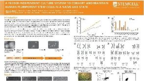 科学海报A Feeder-Independent Culture System to Convert and Maintain Human Pluripotent Stem Cells in a Naïve-Like State
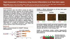 科学海报Rapid Assessment of Pluripotency Using Directed Differentiation to all Three Germ Layers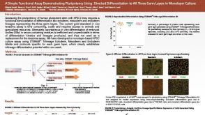

 沪公网安备31010102008431号