MN1 overexpression induces acute myeloid leukemia in mice and predicts ATRA resistance in patients with AML.
Overexpression of wild-type MN1 is a negative prognostic factor in patients with acute myeloid leukemia (AML) with normal cytogenetics. We evaluated whether MN1 plays a functional role in leukemogenesis. We demonstrate using retroviral gene transfer and bone marrow (BM) transplantation that MN1 overexpression rapidly induces lethal AML in mice. Insertional mutagenesis and chromosomal instability were ruled out as secondary aberrations. MN1 increased resistance to all-trans retinoic acid (ATRA)-induced cell-cycle arrest and differentiation by more than 3000-fold in vitro. The differentiation block could be released by fusion of a transcriptional activator (VP16) to MN1 without affecting the ability to immortalize BM cells,suggesting that MN1 blocks differentiation by transcriptional repression. We then evaluated whether MN1 expression levels in patients with AML (excluding M3-AML) correlated with resistance to ATRA treatment in elderly patients uniformly treated within treatment protocol AMLHD98-B. Strikingly,patients with low MN1 expression who received ATRA had a significantly prolonged event-free (P = .008) and overall (P = .04) survival compared with patients with either low MN1 expression and no ATRA,or high MN1 expression with or without ATRA. MN1 is a unique oncogene in hematopoiesis that both promotes proliferation/self-renewal and blocks differentiation,and may become useful as a predictive marker in AML treatment.
View Publication
产品号#:
03234
产品名:
MethoCult™ M3234
Han X-D et al. (MAY 2007)
Proceedings of the National Academy of Sciences of the United States of America 104 21 9007--11
Fetal gene therapy of alpha-thalassemia in a mouse model.
Fetuses with homozygous alpha-thalassemia usually die at the third trimester of pregnancy or soon after birth. Hence,the disease could potentially be a target for fetal gene therapy. We have previously established a mouse model of alpha-thalassemia. These mice mimic the human alpha-thalassemic conditions and can be used as preclinical models for fetal gene therapy. We tested a lentiviral vector containing the HS 2,3,and 4 of the beta-LCR,a central polypurine tract element,and the beta-globin gene promoter directing either the EGFP or the human alpha-globin gene. We showed that the GFP expression was erythroid-specific and detected in BFU-E colonies and the erythroid progenies of CFU-GEMM. For in utero gene delivery,we did yolk sac vessel injection at midgestation of mouse embryos. The recipient mice were analyzed after birth for human alpha-globin gene expression. In the newborn,human alpha-globin gene expression was detected in the liver,spleen,and peripheral blood. The human alpha-globin gene expression was at the peak at 3-4 months,when it reached 20% in some recipients. However,the expression declined at 7 months. Colony-forming assays in these mice showed low abundance of the transduced human alpha-globin gene in their BFU-E and CFU-GEMM and the lack of its transcript. Thus,lentiviral vectors can be an effective vehicle for delivering the human alpha-globin gene into erythroid cells in utero,but,in the mouse model,delivery at late midgestation could not transduce hematopoietic stem cells adequately to sustain gene expression.
View Publication
产品号#:
03434
03444
产品名:
MethoCult™ GF M3434
MethoCult™ GF M3434
North TE et al. (JUN 2007)
Nature 447 7147 1007--11
Haematopoietic stem cell (HSC) homeostasis is tightly controlled by growth factors,signalling molecules and transcription factors. Definitive HSCs derived during embryogenesis in the aorta-gonad-mesonephros region subsequently colonize fetal and adult haematopoietic organs. To identify new modulators of HSC formation and homeostasis,a panel of biologically active compounds was screened for effects on stem cell induction in the zebrafish aorta-gonad-mesonephros region. Here,we show that chemicals that enhance prostaglandin (PG) E2 synthesis increased HSC numbers,and those that block prostaglandin synthesis decreased stem cell numbers. The cyclooxygenases responsible for PGE2 synthesis were required for HSC formation. A stable derivative of PGE2 improved kidney marrow recovery following irradiation injury in the adult zebrafish. In murine embryonic stem cell differentiation assays,PGE2 caused amplification of multipotent progenitors. Furthermore,ex vivo exposure to stabilized PGE2 enhanced spleen colony forming units at day 12 post transplant and increased the frequency of long-term repopulating HSCs present in murine bone marrow after limiting dilution competitive transplantation. The conserved role for PGE2 in the regulation of vertebrate HSC homeostasis indicates that modulation of the prostaglandin pathway may facilitate expansion of HSC number for therapeutic purposes.
View Publication
产品号#:
72192
72194
72372
产品名:
前列腺素E2(Prostaglandin E2)
前列腺素E2(Prostaglandin E2)
16,16-二甲基前列腺素E2
Thein SL et al. (JUL 2007)
Proceedings of the National Academy of Sciences of the United States of America 104 27 11346--51
Intergenic variants of HBS1L-MYB are responsible for a major quantitative trait locus on chromosome 6q23 influencing fetal hemoglobin levels in adults.
Individual variation in fetal hemoglobin (HbF,alpha(2)gamma(2)) response underlies the remarkable diversity in phenotypic severity of sickle cell disease and beta thalassemia. HbF levels and HbF-associated quantitative traits (e.g.,F cell levels) are highly heritable. We have previously mapped a major quantitative trait locus (QTL) controlling F cell levels in an extended Asian-Indian kindred with beta thalassemia to a 1.5-Mb interval on chromosome 6q23,but the causative gene(s) are not known. The QTL encompasses several genes including HBS1L,a member of the GTP-binding protein family that is expressed in erythroid progenitor cells. In this high-resolution association study,we have identified multiple genetic variants within and 5' to HBS1L at 6q23 that are strongly associated with F cell levels in families of Northern European ancestry (P = 10(-75)). The region accounts for 17.6% of the F cell variance in northern Europeans. Although mRNA levels of HBS1L and MYB in erythroid precursors grown in vitro are positively correlated,only HBS1L expression correlates with high F cell alleles. The results support a key role for the HBS1L-related genetic variants in HbF control and illustrate the biological complexity of the mechanism of 6q QTL as a modifier of fetal hemoglobin levels in the beta hemoglobinopathies.
View Publication
产品号#:
09600
09650
产品名:
StemSpan™ SFEM
StemSpan™ SFEM
Dí et al. (DEC 2007)
Cardiovascular research 76 3 517--27
Plasticity of CD133+ cells: role in pulmonary vascular remodeling.
OBJECTIVE: Studies in pulmonary arteries (PA) of patients with chronic obstructive pulmonary disease (COPD) suggest that bone marrow-derived endothelial progenitor cells (CD133(+)) may infiltrate the intima and differentiate into smooth muscle cells (SMC). This study aimed to evaluate the plasticity of CD133(+) cells to differentiate into SMC and endothelial cells (EC) in both cell culture and human isolated PA. METHODS: Plasticity of granulocyte-colony stimulator factor (G-CSF)-mobilized peripheral blood CD133(+) cells was assessed in co-cultures with primary lines of human PA endothelial cells (PAEC) or SMC (PASMC) and in isolated human PA. We also evaluated if the phenotype of differentiated progenitor cells was acquired by fusion or differentiation. RESULTS: The in vitro studies demonstrated CD133(+) cells may acquire the morphology and phenotype of the cells they were co-cultured with. CD133(+) cells co-incubated with human isolated PA were able to migrate into the intima and differentiate into SMC. Progenitor cell differentiation was produced without fusion with mature cells. CONCLUSIONS: We provide evidence of plasticity of CD133(+) cells to differentiate into both endothelial cells and SMC,reinforcing the idea of their potential role in the remodeling process of PA in COPD. This process was conducted by transdifferentiation and not by cell fusion.
View Publication
产品号#:
产品名:
Raffaghello L et al. (JAN 2008)
Stem cells (Dayton,Ohio) 26 1 151--62
Human mesenchymal stem cells inhibit neutrophil apoptosis: a model for neutrophil preservation in the bone marrow niche.
Mesenchymal stem cells (MSC) establish close interactions with bone marrow sinusoids in a putative perivascular niche. These vessels contain a large storage pool of mature nonproliferating neutrophils. Here,we have investigated the effects of human bone marrow MSC on neutrophil survival and effector functions. MSC from healthy donors,at very low MSC:neutrophil ratios (up to 1:500),significantly inhibited apoptosis of resting and interleukin (IL)-8-activated neutrophils and dampened N-formyl-l-methionin-l-leucyl-l-phenylalanine (f-MLP)-induced respiratory burst. The antiapoptotic activity of MSC did not require cell-to-cell contact,as shown by transwell experiments. Antibody neutralization experiments demonstrated that the key MSC-derived soluble factor responsible for neutrophil protection from apoptosis was IL-6,which signaled by activating STAT-3 transcription factor. Furthermore,IL-6 expression was detected in MSC by real-time reverse transcription-polymerase chain reaction and enzyme-linked immunosorbent assay. Finally,recombinant IL-6 was found to protect neutrophils from apoptosis in a dose-dependent manner. MSC had no effect on neutrophil phagocytosis,expression of adhesion molecules,and chemotaxis in response to IL-8,f-MLP,or C5a. These results support the following conclusions: (a) in the bone marrow niche,MSC likely protect neutrophils of the storage pool from apoptosis,preserving their effector functions and preventing the excessive or inappropriate activation of the oxidative metabolism,and (b) a novel mechanism whereby the inflammatory potential of activated neutrophils is harnessed by inhibition of apoptosis and reactive oxygen species production without impairing phagocytosis and chemotaxis has been identified.
View Publication
产品号#:
05401
05402
05411
产品名:
MesenCult™ MSC 基础培养基(人)
MesenCult™ MSC刺激添加物(人)
MesenCult™ 增殖试剂盒(人)
Porayette P et al. (DEC 2007)
Biochemical and biophysical research communications 364 3 522--527
Amyloid-?? precursor protein expression and modulation in human embryonic stem cells: A novel role for human chorionic gonadotropin
The amyloid-beta precursor protein (AbetaPP) is a ubiquitously expressed adhesion and neuritogenic protein whose processing has previously been shown to be regulated by reproductive hormones including the gonadotropin luteinizing hormone (LH) in human neuroblastoma cells. We report for the first time the expression of AbetaPP in human embryonic stem (hES) cells at the mRNA and protein levels. Using N- and C-terminal antibodies against AbetaPP,we detected both the mature and immature forms of AbetaPP as well as truncated variants ( approximately 53kDa,47kDa,and 29kDa) by immunoblot analyses. Expression of AbetaPP is regulated by both the stemness of the cells and pregnancy-associated hormones. Addition of human chorionic gonadotropin,the fetal equivalent of LH that is dramatically elevated during pregnancy,markedly increased the expression of all AbetaPP forms. These results indicate a critical molecular signaling link between the hormonal environment of pregnancy and the expression of AbetaPP in hES cells that is suggestive of an important function for this protein during early human embryogenesis prior to the formation of neural precursor cells.
View Publication
产品号#:
05850
05857
05870
05875
85850
85857
85870
85875
产品名:
mTeSR™1
mTeSR™1
Qiu C et al. (FEB 2008)
Blood 111 4 2400--8
Globin switches in yolk sac-like primitive and fetal-like definitive red blood cells produced from human embryonic stem cells.
We have previously shown that coculture of human embryonic stem cells (hESCs) for 14 days with immortalized fetal hepatocytes yields CD34(+) cells that can be expanded in serum-free liquid culture into large numbers of megaloblastic nucleated erythroblasts resembling yolk sac-derived cells. We show here that these primitive erythroblasts undergo a switch in hemoglobin (Hb) composition during late terminal erythroid maturation with the basophilic erythroblasts expressing predominantly Hb Gower I (zeta(2)epsilon(2)) and the orthochromatic erythroblasts hemoglobin Gower II (alpha(2)epsilon(2)). This suggests that the switch from Hb Gower I to Hb Gower II,the first hemoglobin switch in humans is a maturation switch not a lineage switch. We also show that extending the coculture of the hESCs with immortalized fetal hepatocytes to 35 days yields CD34(+) cells that differentiate into more developmentally mature,fetal liver-like erythroblasts,that are smaller,express mostly fetal hemoglobin,and can enucleate. We conclude that hESC-derived erythropoiesis closely mimics early human development because the first 2 human hemoglobin switches are recapitulated,and because yolk sac-like and fetal liver-like cells are sequentially produced. Development of a method that yields erythroid cells with an adult phenotype remains necessary,because the most mature cells that can be produced with current systems express less than 2% adult beta-globin mRNA.
View Publication
产品号#:
09600
09650
18056
18056RF
产品名:
StemSpan™ SFEM
StemSpan™ SFEM
Ghiaur G et al. (APR 2008)
Blood 111 7 3313--21
Rac1 is essential for intraembryonic hematopoiesis and for the initial seeding of fetal liver with definitive hematopoietic progenitor cells.
Definitive hematopoietic stem and progenitor cells (HSCs/Ps) originating from the yolk sac and/or para-aorta-splanchno-pleura/aorta-gonad-mesonephros are hypothesized to colonize the fetal liver,but mechanisms involved are poorly defined. The Rac subfamily of Rho GTPases has been shown to play essential roles in HSC/P localization to the bone marrow following transplantation. Here,we study the role of Rac1 in HSC/P migration during ontogeny and seeding of fetal liver. Using a triple-transgenic approach,we have deleted Rac1 in HSCs/Ps during very early embryonic development. Without Rac1,there was a decrease in circulating HSCs/Ps in the blood of embryonic day (E) 10.5 embryos,while yolk sac definitive hematopoiesis was quantitatively normal. Intraembryonic hematopoiesis was significantly impaired in Rac1-deficient embryos,culminating with absence of intra-aortic clusters and fetal liver hematopoiesis. At E10.5,Rac1-deficient HSCs/Ps displayed decreased transwell migration and impaired inter-action with the microenvironment in migration-dependent assays. These data suggest that Rac1 plays an important role in HSC/P migration during embryonic development and is essential for the emergence of intraembryonic hematopoiesis.
View Publication


 EasySep™小鼠TIL(CD45)正选试剂盒




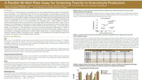 科学海报A Flexible 96-Well Plate Assay for Screening Toxicity to Granulocyte Production
 沪公网安备31010102008431号