
 EasySep™小鼠TIL(CD45)正选试剂盒


技术资料
-
-
-
-
技术窍门从脐带血中分离CD34+细胞
-
-
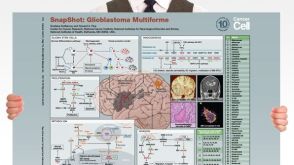 挂图SnapShot: Glioblastoma Multiforme Overview of the key concepts and mechanisms in glioblastoma multiforme biology


 沪公网安备31010102008431号