The differentiation of eosinophils from hematopoietic precursors and their subsequent maturation,chemotaxis,and activation is primarily regulated by interleukin-5 (IL-5). To examine the effect of chronic IL-5 exposure on hematopoiesis,IL-5 transgenic (IL-5trg) mice and wild-type BALB/c (WT) mice were examined. In comparison to WT mice,a significant alteration in bone marrow hematopoiesis was observed in IL-5trg mice. Although the total number of myeloid progenitors in the bone marrow of IL-5trg mice was not significantly altered,the number of long-term culture-initiating cells (LTC-ICs) was 1.5-fold lower than that observed in WT mice. Furthermore,IL-5trg mice failed to demonstrate hematopoietic activity in long-term bone marrow cultures,which correlated with a significant decrease in the number of bone marrow mesenchymal/stromal progenitor (MSP) cells in these mice. In comparison to WT mice,a 10-fold decrease was observed in the number of fibroblast colony-forming units (CFU-Fs) in IL-5trg bone marrow. Hematopoietic activity of IL-5trg bone marrow cells was rescued by cultivation on preestablished layers of bone marrow-derived stromal cells. However,in contrast to bone marrow,increased hematopoietic activity was observed in the spleen and peripheral blood of IL-5trg mice. Likewise,the numbers of LTC-ICs and granulocyte-macrophage,macrophage,eosinophil,B-lymphocyte progenitors in the peripheral blood and spleen of IL-5trg mice were approximately 20-fold higher than in WT mice. A significant increase in CFU-F numbers was also observed in the spleens of IL-5trg mice compared with WT mice. Overall,our results suggest that constitutive overexpression of IL-5 can potentially induce colonization of spleen with MSP cells,which provides the necessary microenvironment for establishment of hematopoiesis in extramedullary sites.
View Publication

 EasySep™小鼠TIL(CD45)正选试剂盒


 技术窍门组织解离方法指南
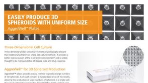
 挂图The Identity and Properties of Mesenchymal Stem Cells Overview of MSC expansion, differentiation, immunoregulatory properties and therapeutic potential


 沪公网安备31010102008431号