The monoclonal antibody 97A6 defines a novel surface antigen expressed on human basophils and their multipotent and unipotent progenitors.
Basophils (Ba) and mast cells (MC) are important effector cells of inflammatory reactions. Both cell types derive from CD34(+) hematopoietic progenitors. However,little is known about the cell subsets that become committed to and give rise to Ba and/or MC. We have generated a monoclonal antibody (MoAb),97A6,that specifically detects human Ba,MC (lung,skin),and their CD34(+) progenitors. Other mature hematopoietic cells (neutrophils,eosinophils,monocytes,lymphocytes,platelets) did not react with MoAb 97A6,and sorting of 97A6(+) peripheral blood (PB) and bone marrow (BM) cells resulted in an almost pure population (textgreater98%) of Ba. Approximately 1% of CD34(+) BM and PB cells was found to be 97A6(+). Culture of sorted CD34(+)97A6(+) BM cells in semisolid medium containing phytohemagglutinin-stimulated leukocyte supernatant for 16 days (multilineage assay) resulted in the formation of pure Ba colonies (10 of 40),Ba-eosinophil colonies (7 of 40),Ba-macrophage colonies (3 of 40),and multilineage Ba-eosinophil-macrophage and/or neutrophil colonies (12 of 40). In contrast,no Ba could be cultured from CD34(+)97A6(-) cells. Liquid culture of CD34(+) PB cells in the presence of 100 ng/mL interleukin (IL)-3 (Ba progenitor assay) resulted in an increase of 97A6(+) cells,starting from 1% of day-0 cells to almost 70% (basophils) after day 7. Culture of sorted BM CD34(+)97A6(+) cells in the presence of 100 ng/mL stem cell factor (SCF) for 35 days (mast cell progenitor assay) resulted in the growth of MC (textgreater30% on day 35). Anti-IgE-induced IgE receptor cross-linking on Ba for 15 minutes resulted in a 4-fold to 5-fold upregulation of 97A6 antigen expression. These data show that the 97A6-reactive antigen plays a role in basophil activation and is expressed on multipotent CD34(+) progenitors,MC progenitors,Ba progenitors,as well as on mature Ba and tissue MC. The lineage-specificity of MoAb 97A6 suggests that this novel marker may be a useful tool to isolate and analyze Ba/MC and their progenitors.
View Publication
Kunishima S et al. (MAR 2008)
Blood 111 6 3015--23
Differential expression of wild-type and mutant NMMHC-IIA polypeptides in blood cells suggests cell-specific regulation mechanisms in MYH9 disorders.
MYH9 disorders such as May-Hegglin anomaly are characterized by macrothrombocytopenia and cytoplasmic granulocyte inclusion bodies that result from mutations in MYH9,the gene for nonmuscle myosin heavy chain-IIA (NMMHC-IIA). We examined the expression of mutant NMMHC-IIA polypeptide in peripheral blood cells from patients with MYH9 5770delG and 5818delG mutations. A specific antibody to mutant NMMHC-IIA (NT629) was raised against the abnormal carboxyl-terminal residues generated by 5818delG. NT629 reacted to recombinant 5818delG NMMHC-IIA but not to wild-type NMMHC-IIA,and did not recognize any cellular components of normal peripheral blood cells. Immunofluorescence and immunoblotting revealed that mutant NMMHC-IIA was present and sequestrated only in inclusion bodies within neutrophils,diffusely distributed throughout lymphocyte cytoplasm,sparsely localized on a diffuse cytoplasmic background in monocytes,and uniformly distributed at diminished levels only in large platelets. Mutant NMMHC-IIA did not translocate to lamellipodia in surface activated platelets. Wild-type NMMHC-IIA was homogeneously distributed among megakaryocytes derived from the peripheral blood CD34(+) cells of patients,but coarse mutant NMMHC-IIA was heterogeneously scattered without abnormal aggregates in the cytoplasm. We show the differential expression of mutant NMMHC-IIA and postulate that cell-specific regulation mechanisms function in MYH9 disorders.
View Publication
产品号#:
09600
09650
产品名:
StemSpan™ SFEM
StemSpan™ SFEM
Elling C et al. (MAR 2011)
Blood 117 10 2935--43
Novel imatinib-sensitive PDGFRA-activating point mutations in hypereosinophilic syndrome induce growth factor independence and leukemia-like disease.
The FIP1L1-PDGFRA fusion is seen in a fraction of cases with a presumptive diagnosis of hypereosinophilic syndrome (HES). However,because most HES patients lack FIP1L1-PDGFRA,we studied whether they harbor activating mutations of the PDGFRA gene. Sequencing of 87 FIP1L1-PDGFRA-negative HES patients revealed several novel PDGFRA point mutations (R481G,L507P,I562M,H570R,H650Q,N659S,L705P,R748G,and Y849S). When cloned into 32D cells,N659S and Y849S and-on selection for high expressors-also H650Q and R748G mutants induced growth factor-independent proliferation,clonogenic growth,and constitutive phosphorylation of PDGFRA and Stat5. Imatinib antagonized Stat5 phosphorylation. Mutations involving positions 659 and 849 had been shown previously to possess transforming potential in gastrointestinal stromal tumors. Because H650Q and R748G mutants possessed only weak transforming activity,we injected 32D cells harboring these mutants or FIP1L1-PDGFRA into mice and found that they induced a leukemia-like disease. Oral imatinib treatment significantly decreased leukemic growth in vivo and prolonged survival. In conclusion,our data provide evidence that imatinib-sensitive PDGFRA point mutations play an important role in the pathogenesis of HES and we propose that more research should be performed to further define the frequency and treatment response of PDGFRA mutations in FIP1L1-PDGFRA-negative HES patients.
View Publication
产品号#:
03231
产品名:
MethoCult™ M3231
Kim M-H et al. (MAR 2011)
Blood 117 12 3343--52
Neutrophil survival and c-kit(+)-progenitor proliferation in Staphylococcus aureus-infected skin wounds promote resolution.
Polymorphonuclear neutrophils (PMNs) are critical for the formation,maintenance,and resolution of bacterial abscesses. However,the mechanisms that regulate PMN survival and proliferation during the evolution of an abscess are not well defined. Using a mouse model of Staphylococcus aureus abscess formation within a cutaneous wound,combined with real-time imaging of genetically tagged PMNs,we observed that a high bacterial burden elicited a sustained mobilization of PMNs from the bone marrow to the infected wound,where their lifespan was markedly extended. A continuous rise in wound PMN number,which was not accounted for by trafficking from the bone marrow or by prolonged survival,was correlated with the homing of c-kit(+)-progenitor cells from the blood to the wound,where they proliferated and formed mature PMNs. Furthermore,by blocking their recruitment with an antibody to c-kit,which severely limited the proliferation of mature PMNs in the wound and shortened mouse survival,we confirmed that progenitor cells are not only important contributors to PMN expansion in the wound,but are also functionally important for immune protection. We conclude that the abscess environment provides a niche capable of regulating PMN survival and local proliferation of bone marrow-derived c-kit(+)-progenitor cells.
View Publication
产品号#:
03434
03444
产品名:
MethoCult™ GF M3434
MethoCult™ GF M3434
Eash KJ et al. (MAY 2009)
Blood 113 19 4711--9
CXCR4 is a key regulator of neutrophil release from the bone marrow under basal and stress granulopoiesis conditions.
The number of neutrophils in the blood is tightly regulated to ensure adequate protection against microbial pathogens while minimizing damage to host tissue. Neutrophil homeostasis in the blood is achieved through a balance of neutrophil production,release from the bone marrow,and clearance from the circulation. Accumulating evidence suggests that signaling by CXCL12,through its major receptor CXCR4,plays a key role in maintaining neutrophil homeostasis. Herein,we generated mice with a myeloid lineage-restricted deletion of CXCR4 to define the mechanisms by which CXCR4 signals regulate this process. We show that CXCR4 negatively regulates neutrophil release from the bone marrow in a cell-autonomous fashion. However,CXCR4 is dispensable for neutrophil clearance from the circulation. Neutrophil mobilization responses to granulocyte colony-stimulating factor (G-CSF),CXCL2,or Listeria monocytogenes infection are absent or impaired,suggesting that disruption of CXCR4 signaling may be a common step mediating neutrophil release. Collectively,these data suggest that CXCR4 signaling maintains neutrophil homeostasis in the blood under both basal and stress granulopoiesis conditions primarily by regulating neutrophil release from the bone marrow.
View Publication
产品号#:
03434
03444
产品名:
MethoCult™ GF M3434
MethoCult™ GF M3434
Velu CS et al. (MAY 2009)
Blood 113 19 4720--8
Gfi1 regulates miR-21 and miR-196b to control myelopoiesis.
The zinc finger protein growth factor independent-1 (Gfi1) is a transcriptional repressor that is critically required for normal granulocytic differentiation. GFI1 loss-of-function mutations are found in some patients with severe congenital neutropenia (SCN). The SCN-associated GFI1-mutant proteins act as dominant negatives to block granulopoiesis through selective deregulation of a subset of GFI1 target genes. Here we show that Gfi1 is a master regulator of microRNAs,and that deregulated expression of these microRNAs recapitulates a Gfi1 loss-of-function block to granulocyte colony-stimulating factor (G-CSF)-stimulated granulopoiesis. Specifically,bone marrow cells from a GFI1-mutant SCN patient and Gfi1(-/-) mice display deregulated expression of miR-21 and miR-196B expression. Flow cytometric analysis and colony assays reveal that the overexpression or depletion of either miR induces changes in myeloid development. However,coexpression of miR-21 and miR-196b (as seen in Gfi1(-/-) mice and a GFI1N382S SCN patient) completely blocks G-CSF-induced granulopoiesis. Thus,our results not only identify microRNAs whose regulation is required during myelopoiesis,but also provide an example of synergy in microRNA biologic activity and illustrate potential mechanisms underlying SCN disease pathogenesis.
View Publication
产品号#:
03534
09600
09650
产品名:
MethoCult™ GF M3534
StemSpan™ SFEM
StemSpan™ SFEM
Dutt S et al. (MAR 2011)
Blood 117 9 2567--76
Haploinsufficiency for ribosomal protein genes causes selective activation of p53 in human erythroid progenitor cells.
Haploinsufficiency for ribosomal protein genes has been implicated in the pathophysiology of Diamond-Blackfan anemia (DBA) and the 5q-syndrome,a subtype of myelodysplastic syndrome. The p53 pathway is activated by ribosome dysfunction,but the molecular basis for selective impairment of the erythroid lineage in disorders of ribosome function has not been determined. We found that p53 accumulates selectively in the erythroid lineage in primary human hematopoietic progenitor cells after expression of shRNAs targeting RPS14,the ribosomal protein gene deleted in the 5q-syndrome,or RPS19,the most commonly mutated gene in DBA. Induction of p53 led to lineage-specific accumulation of p21 and consequent cell cycle arrest in erythroid progenitor cells. Pharmacologic inhibition of p53 rescued the erythroid defect,whereas nutlin-3,a compound that activates p53 through inhibition of HDM2,selectively impaired erythropoiesis. In bone marrow biopsies from patients with DBA or del(5q) myelodysplastic syndrome,we found an accumulation of nuclear p53 staining in erythroid progenitor cells that was not present in control samples. Our findings indicate that the erythroid lineage has a low threshold for the induction of p53,providing a basis for the failure of erythropoiesis in the 5q-syndrome,DBA,and perhaps other bone marrow failure syndromes.
View Publication
产品号#:
03334
03434
03444
产品名:
MethoCult™ M3334
MethoCult™ GF M3434
MethoCult™ GF M3434
Simons MP et al. (MAR 2008)
Journal of leukocyte biology 83 3 621--9
TNF-related apoptosis-inducing ligand (TRAIL) is expressed throughout myeloid development, resulting in a broad distribution among neutrophil granules.
TRAIL induces apoptosis in a variety of tumor cells. Our laboratory found that human neutrophils contain an intracellular reservoir of prefabricated TRAIL that is released after stimulation with Mycobacterium bovis bacillus Calmette-Guérin. In this study,we examined the subcellular distribution of TRAIL in freshly isolated neutrophils. Neutrophil granules,secretory vesicles (SV),and plasma membrane vesicles were isolated by subcellular fractionation,followed by free-flow electrophoresis,and examined by ELISA and immunoblot. TRAIL was found in all membrane-bound fractions with the highest amounts in the fractions enriched in azurophilic granule (AG) and SV. Immunofluorescence confocal microscopy showed that TRAIL colocalized independently with myeloperoxidase (MPO),lactoferrin (LF),and albumin,respective markers of AG,specific granules,and SV. Furthermore,immunotransmission electron microscopy demonstrated that TRAIL colocalized intracellularly with MPO and albumin. We examined TRAIL expression in PLB-985 cells induced with dimethylformamide and in CD34-positive stem cells treated with G-CSF. Quantitative RT-PCR analysis showed that TRAIL was expressed in each stage of development,whereas MPO and LF were only expressed at distinct times during differentiation. Collectively,these findings suggest that TRAIL is expressed throughout neutrophil development,resulting in a broad distribution among different granule subtypes.
View Publication
产品号#:
09600
09650
09850
产品名:
StemSpan™ SFEM
StemSpan™ SFEM
El-Ouriaghli F et al. (NOV 2003)
Blood 102 10 3786--92
Clonal dominance of chronic myelogenous leukemia is associated with diminished sensitivity to the antiproliferative effects of neutrophil elastase.
Clinical observations suggest that in chronic myelogenous leukemia (CML),the Philadelphia chromosome (Ph+) clone has a growth advantage over normal hematopoiesis. Patients with CML have high levels of neutrophil elastase,which has recently been shown to antagonize the action of granulocyte-colony-stimulating factor (G-CSF) and other growth factors. We therefore compared the effect of elastase on the growth of normal and CML progenitor cells. In 10-day suspension cultures of normal or CML CD34+ cells supplemented with G-CSF,stem cell factor (SCF),and granulocyte macrophage-colony-stimulating factor (GM-CSF),CML cells had diminished sensitivity to the growth inhibitory effect of elastase. When equal numbers of CML and normal CD34+ cells were cocultured for 10 days,there was no change in the relative proportions of normal and leukemic cells (measured by fluorescence in situ hybridization [FISH] or flow cytometry). However,when elastase was added,CML cells predominated at the end of the culture period (78% vs 22% with 1 microg/mL and 80% vs 20% with 5 microg/mL elastase). CML neutrophils substituted effectively for elastase in suppressing the proliferation of normal CD34+ cells,but this effect was abrogated by serine protease inhibitors. These results suggest that elastase overproduction by the leukemic clone can change the growth environment by digesting growth factors,thereby giving advantage to Ph+ hematopoiesis.
View Publication

 EasySep™小鼠TIL(CD45)正选试剂盒


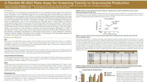 科学海报A Flexible 96-Well Plate Assay for Screening Toxicity to Granulocyte Production

 沪公网安备31010102008431号