Hwang GH et al. (DEC 2017)
Journal of cellular physiology 232 12 3384--3395
Purification of small molecule-induced cardiomyocytes from human induced pluripotent stem cells using a reporter system.
In order to realize the practical use of human pluripotent stem cell (hPSC)-derived cardiomyocytes for the purpose of clinical use or cardiovascular research,the generation of large numbers of highly purified cardiomyocytes should be achieved. Here,we show an efficient method for cardiac differentiation of human induced pluripotent stem cells (hiPSCs) in chemically defined conditions and purification of hiPSC-derived cardiomyocytes using a reporter system. Regulation of the Wnt/β-catenin signaling pathway is implicated in the induction of the cardiac differentiation of hPSCs. We increased cardiac differentiation efficiency of hiPSCs in chemically defined conditions through combined treatment with XAV939,a tankyrase inhibitor and IWP2,a porcupine inhibitor and optimized concentrations. Although cardiac differentiation efficiency was high (>80%),it was difficult to suppress differentiation into non-cardiac cells,Therefore,we applied a lentiviral reporter system,wherein green fluorescence protein (GFP) and Zeocin-resistant gene are driven by promoter activation of a gene (TNNT2) encoding cardiac troponin T (cTnT),a cardiac-specific protein,to exclude non-cardiomyocytes from differentiated cell populations. We transduced this reporter construct into differentiated cells using a lentiviral vector and then obtained highly purified hiPSC-derived cardiomyocytes by treatment with the lowest effective dose of Zeocin. We significantly increased transgenic efficiency through manipulation of the cells in which the differentiated cells were simultaneously infected with virus and re-plated after single-cell dissociation. Purified cells specifically expressed GFP,cTnT,displayed typical properties of cardiomyocytes. This study provides an efficient strategy for obtaining large quantities of highly purified hPSC-derived cardiomyocytes for application in regenerative medicine and biomedical research.
View Publication
产品号#:
85850
85857
85870
85875
产品名:
mTeSR™1
mTeSR™1
Ware CB et al. (MAR 2014)
Proceedings of the National Academy of Sciences of the United States of America 111 12 4484--9
Derivation of naive human embryonic stem cells.
The naïve pluripotent state has been shown in mice to lead to broad and more robust developmental potential relative to primed mouse epiblast cells. The human naïve ES cell state has eluded derivation without the use of transgenes,and forced expression of OCT4,KLF4,and KLF2 allows maintenance of human cells in a naïve state [Hanna J,et al. (2010) Proc Natl Acad Sci USA 107(20):9222-9227]. We describe two routes to generate nontransgenic naïve human ES cells (hESCs). The first is by reverse toggling of preexisting primed hESC lines by preculture in the histone deacetylase inhibitors butyrate and suberoylanilide hydroxamic acid,followed by culture in MEK/ERK and GSK3 inhibitors (2i) with FGF2. The second route is by direct derivation from a human embryo in 2i with FGF2. We show that human naïve cells meet mouse criteria for the naïve state by growth characteristics,antibody labeling profile,gene expression,X-inactivation profile,mitochondrial morphology,microRNA profile and development in the context of teratomas. hESCs can exist in a naïve state without the need for transgenes. Direct derivation is an elusive,but attainable,process,leading to cells at the earliest stage of in vitro pluripotency described for humans. Reverse toggling of primed cells to naïve is efficient and reproducible.
View Publication
产品号#:
05860
05880
产品名:
Cheung C et al. (APR 2014)
Nature protocols 9 4 929--38
Directed differentiation of embryonic origin-specific vascular smooth muscle subtypes from human pluripotent stem cells.
Vascular smooth muscle cells (SMCs) arise from diverse developmental origins. Regional distribution of vascular diseases may,in part,be attributed to this inherent heterogeneity in SMC lineage. Therefore,systems for generating human SMC subtypes of distinct embryonic origins would represent useful platforms for studying the influence of SMC lineage on the spatial specificity of vascular disease. Here we describe how human pluripotent stem cells can be differentiated into distinct populations of SMC subtypes under chemically defined conditions. The initial stage (days 0-5 or 0-7) begins with the induction of three intermediate lineages: neuroectoderm,lateral plate mesoderm and paraxial mesoderm. Subsequently,these precursor lineages are differentiated into contractile SMCs (days 5-19+). At key stages,the emergence of lineage-specific markers confirms recapitulation of embryonic developmental pathways and generation of functionally distinct SMC subtypes. The ability to derive an unlimited supply of human SMCs will accelerate applications in regenerative medicine and disease modeling.
View Publication
产品号#:
05850
05857
05870
05875
85850
85857
85870
85875
产品名:
mTeSR™1
mTeSR™1
Lam AT-L et al. (JUL 2014)
Stem cells and development 23 14 1688--1703
Cationic Surface Charge Combined with Either Vitronectin or Laminin Dictates the Evolution of Human Embryonic Stem Cells/Microcarrier Aggregates and Cell Growth in Agitated Cultures
The expansion of human pluripotent stem cells (hPSC) for biomedical applications generally compels a defined,reliable,and scalable platform. Bioreactors offer a three-dimensional culture environment that relies on the implementation of microcarriers (MC),as supports for cell anchorage and their subsequent growth. Polystyrene microspheres/MC coated with adhesion-promoting extracellular matrix (ECM) protein,vitronectin (VN),or laminin (LN) have been shown to support hPSC expansion in a static environment. However,they are insufficient to promote human embryonic stem cells (hESC) seeding and their expansion in an agitated environment. The present study describes an innovative technology,consisting of a cationic charge that underlies the ECM coatings. By combining poly-L-lysine (PLL) with a coating of ECM protein,cell attachment efficiency and cell spreading are improved,thus enabling seeding under agitation in a serum-free medium. This coating combination also critically enables the subsequent formation and evolution of hPSC/MC aggregates,which ensure cell viability and generate high yields. Aggregate dimensions of at least 300 $\$ during early cell growth give rise to ≈15-fold expansion at 7 days' culture. Increasing aggregate numbers at a quasi-constant size of ≈300 $\$ indicates hESC growth within a self-regulating microenvironment. PLL+LN enables cell seeding and aggregate evolution under constant agitation,whereas PLL+VN requires an intermediate 2-day static pause to attain comparable aggregate sizes and correspondingly high expansion yields. The cells' highly reproducible bioresponse to these defined and characterized MC surface properties is universal across multiple cell lines,thus confirming the robustness of this scalable expansion process in a defined environment.
View Publication
产品号#:
05850
05857
05870
05875
85850
85857
85870
85875
产品名:
mTeSR™1
mTeSR™1
ndrea de Oliveira Georges JA et al. (AUG 2014)
Stem cell reviews 10 4 472--479
Aberrant patterns of X chromosome inactivation in a new line of human embryonic stem cells established in physiological oxygen concentrations
One of the differences between murine and human embryonic stem cells (ESCs) is the epigenetic state of the X chromosomes in female lines. Murine ESCs (mESCs) present two transcriptionally active Xs that will undergo the dosage compensation process of XCI upon differentiation,whereas most human ESCs (hESCs) spontaneously inactivate one X while keeping their pluripotency. Whether this reflects differences in embryonic development of mice and humans,or distinct culture requirements for the two kinds of pluripotent cells is not known. Recently it has been shown that hESCs established in physiological oxygen levels are in a stable pre-XCI state equivalent to that of mESCs,suggesting that culture in low oxygen concentration is enough to preserve that epigenetic state of the X chromosomes. Here we describe the establishment of two new lines of hESCs under physiological oxygen level and the characterization of the XCI state in the 46,XX line BR-5. We show that a fraction of undifferentiated cells present XIST RNA accumulation and single H3K27me foci,characteristic of the inactive X. Moreover,analysis of allele specific gene expression suggests that pluripotent BR-5 cells present completely skewed XCI. Our data indicate that physiological levels of oxygen are not sufficient for the stabilization of the pre-XCI state in hESCs.
View Publication
产品号#:
05850
05857
05870
05875
85850
85857
85870
85875
产品名:
mTeSR™1
mTeSR™1
Brzeszczynska J et al. (JUN 2014)
International journal of molecular medicine 33 6 1597--1606
Differentiation and molecular profiling of human embryonic stem cell-derived corneal epithelial cells
It has been suggested that the isolation of scalable populations of limbal stem cells may lead to radical changes in ocular therapy. In particular,the derivation and transplantation of corneal stem cells from these populations may result in therapies providing clinical normality of the diseased or damaged cornea. Although feasible in theory,the lack of donor material in sufficient quantity and quality currently limits such a strategy. A potential scalable source of corneal cells could be derived from pluripotent stem cells (PSCs). We developed an in vitro and serum-free corneal differentiation model which displays significant promise. Our stepwise differentiation model was designed with reference to development and gave rise to cells which displayed similarities to epithelial progenitor cells which can be specified to cells displaying a corneal epithelial phenotype. We believe our approach is novel,provides a robust model of human development and in the future,may facilitate the generation of corneal epithelial cells that are suitable for clinical use. Additionally,we demonstrate that following continued cell culture,stem cell-derived corneal epithelial cells undergo transdifferentiation and exhibit squamous metaplasia and therefore,also offer an in vitro model of disease.
View Publication
产品号#:
05850
05857
05870
05875
85850
85857
85870
85875
产品名:
mTeSR™1
mTeSR™1
Risau W et al. (MAR 1988)
Development (Cambridge,England) 102 3 471--8
Vasculogenesis and angiogenesis in embryonic-stem-cell-derived embryoid bodies.
Embryonic stem cells (ESC) have been established previously from the inner cell mass cells of mouse blastocysts. In suspension culture,they spontaneously differentiate to blood-island-containing cystic embryoid bodies (CEB). The development of blood vessels from in situ differentiating endothelial cells of blood islands,a process which we call vasculogenesis,was induced by injecting ESC into the peritoneal cavity of syngeneic mice. In the peritoneum,fusion of blood islands and formation of an in vivo-like primary capillary plexus occurred. Transplantation of ESC and ESC-derived complex and cystic embryoid bodies (ESC-CEB) onto the quail chorioallantoic membrane (CAM) induced an angiogenic response,which was directed by nonyolk sac endoderm structures. Neither yolk sac endoderm from ESC-CEB nor normal mouse yolk sac tissue induced angiogenesis on the quail CAM. Extracts from ESC-CEB stimulated the proliferation of capillary endothelial cells in vitro. Mitogenic activity increase during in vitro culture and differentiation of ESC. Almost all growth factor activity was associated with the cells. The ESC-CEB derived endothelial cell growth factor bound to heparin-sepharose. The identification of acidic fibroblast growth factor (FGF)in heparin-sepharose-purified material was accomplished by immunoblot experiments involving antibodies against acidic and basic FGF. We conclude that vasculogenesis,the development of blood vessels from in situ differentiating endothelial cells,and angiogenesis,the sprouting of capillaries from preexisting vessels are very early events during embryogenesis which can be studied using ESC differentiating in vitro. Our results suggest that vasculogenesis and angiogenesis are differently regulated.
View Publication
产品号#:
06902
06952
00321
00322
00323
00324
00325
产品名:
Yang W-T and Zheng P-S (FEB 2014)
PloS one 9 2 e88827
Promoter hypermethylation of KLF4 inactivates its tumor suppressor function in cervical carcinogenesis.
OBJECTIVE The KLF4 gene has been shown to be inactivated in cervical carcinogenesis as a tumor suppressor. However,the mechanism of KLF4 silencing in cervical carcinomas has not yet been identified. DNA methylation plays a key role in stable suppression of gene expression. METHODS The methylation status of the KLF4 promoter CpG islands was analyzed by bisulfite sequencing (BSQ) in tissues of normal cervix and cervical cancer. KLF4 gene expression was detected by RT-PCR,immunohistochemistry and western blot. KLF4 promoter methylation in cervical cancer cell line was determined by BSQ and methylation-specific polymerase chain reaction (MS-PCR). Cell proliferation ability was detected by cell growth curve and MTT assay. RESULTS The methylated allele was found in 41.90% of 24 cervical cancer tissues but only in 11.11% of 11 normal cervix tissues (Ptextless0.005). KLF4 mRNA levels were significantly reduced in cervical cancer tissues compared with normal cervix tissues (Ptextless0.01) and KLF4 mRNA expression showed a significant negative correlation with the promoter hypermethylation (r = -0.486,P = 0.003). Cervical cancer cell lines also showed a significant negative correlation between KLF4 expression and hypermethylation. After treatment with the demethylating agent 5-Azacytidine (5-Aza),the expression of KLF4 in the cervical cancer cell lines at both mRNA and protein levels was drastically increased,the cell proliferation ability was inhibited and the chemosensitivity for cisplatin was significantly increased. CONCLUSION KLF4 gene is inactivated by methylation-induced silencing mechanisms in a large subset of cervical carcinomas and KLF4 promoter hypermethylation inactivates the gene's function as a tumor suppressor in cervical carcinogenesis.
View Publication


 EasySep™小鼠TIL(CD45)正选试剂盒





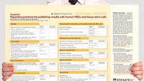 挂图Reporting Practices for Publishing Results with hPSCs Learn how to plan and conduct your human pluripotent stem cell (hPSC)-based research following the ISSCR’s Standards for Human Stem Cell Use in Research
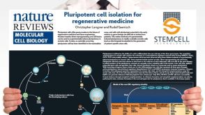 挂图Pluripotent Stem Cell Biology Nuclear transfer, reprogramming and cell fusion to induce pluripotency in somatic cells
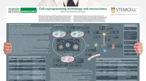 挂图Cell-Reprogramming Technology and Neuroscience Details on human iPSC-derived models of neuropsychiatric and neurodegenerative disorders
 沪公网安备31010102008431号