Xie X et al. (JAN 2011)
Stem cells and development 20 1 127--138
Effects of long-term culture on human embryonic stem cell aging.
In recent years,human embryonic stem (hES) cells have become a promising cell source for regenerative medicine. Although hES cells have the ability for unlimited self-renewal,potential adverse effects of long-term cell culture upon hES cells must be investigated before therapeutic applications of hES cells can be realized. Here we investigated changes in molecular profiles associated with young (textless60 passages) and old (textgreater120 passages) cells of the H9 hES cell line as well as young (textless85 passages) and old (textgreater120 passages) cells of the PKU1 hES cell line. Our results show that morphology,stem cell markers,and telomerase activity do not differ significantly between young and old passage cells. Cells from both age groups were also shown to differentiate into derivatives of all 3 germ layers upon spontaneous differentiation in vitro. Interestingly,mitochondrial dysfunction was found to occur with prolonged culture. Old passage cells of both the H9 and PKU1 lines were characterized by higher mitochondrial membrane potential,larger mitochondrial morphology,and higher reactive oxygen species content than their younger counterparts. Teratomas derived from higher passage cells were also found to have an uneven preference for differentiation compared with tumors derived from younger cells. These findings suggest that prolonged culture of hES cells may negatively impact mitochondrial function and possibly affect long-term pluripotency.
View Publication
产品号#:
05850
05857
05870
05875
85850
85857
85870
85875
产品名:
mTeSR™1
mTeSR™1
Fraga AM et al. (MAR 2011)
Cell Transplantation 20 3 431--40
Establishment of a Brazilian line of human embryonic stem cells in defined medium: implications for cell therapy in an ethnically diverse population.
Pluripotent human embryonic stem (hES) cells are an important experimental tool for basic and applied research,and a potential source of different tissues for transplantation. However,one important challenge for the clinical use of these cells is the issue of immunocompatibility,which may be dealt with by the establishment of hES cell banks to attend different populations. Here we describe the derivation and characterization of a line of hES cells from the Brazilian population,named BR-1,in commercial defined medium. In contrast to the other hES cell lines established in defined medium,BR-1 maintained a stable normal karyotype as determined by genomic array analysis after 6 months in continuous culture (passage 29). To our knowledge,this is the first reported line of hES cells derived in South America. We have determined its genomic ancestry and compared the HLA-profile of BR-1 and another 22 hES cell lines established elsewhere with those of the Brazilian population,finding they would match only 0.011% of those individuals. Our results highlight the challenges involved in hES cell banking for populations with a high degree of ethnic admixture.
View Publication
产品号#:
05850
05857
05870
05875
85850
85857
85870
85875
产品名:
mTeSR™1
mTeSR™1
Leung HW et al. (FEB 2011)
Tissue engineering. Part C,Methods 17 2 165--72
Agitation can induce differentiation of human pluripotent stem cells in microcarrier cultures.
One of the factors that can impact human embryonic stem cell expansion in stirred microcarrier culture reactors is mechanical stress caused by agitation. Therefore,we have investigated the effects of agitation on human embryonic stem cell growth and expression of pluripotent markers. Agitation of HES-2 cell line in microcarrier cultures in stirred spinner and agitated six-well plates did not affect expression of pluripotent markers,cell viability,and cell doubling times even after seven passages. However,HES-3 cell line was found to be shear sensitive,showing downregulation of three pluripotent markers Oct-4,mAb 84,and Tra-1-60,and lower cell densities in agitated as compared with static cultures,even after one passage. Cell viability was unaffected. The HES-3-agitated cultures showed increased expression of genes and proteins of the three germ layers. We were unable to prevent loss of pluripotent markers or restore doubling times in agitated HES-3 microcarrier cultures by addition of five different known cell protective polymers. In addition,the human induced pluripotent cell line IMR90 was also shown to differentiate in agitated conditions. These results indicate that the effect of agitation on cell growth and differentiation is cell line specific. We assume that the changes in the growth and differentiation of the agitation-sensitive (HES-3) cell line do not result from the effect of shear stress directly on cell viability,but rather by signaling effects that influence the cells to differentiate resulting in slower growth.
View Publication
产品号#:
05850
05857
05870
05875
85850
85857
85870
85875
产品名:
mTeSR™1
mTeSR™1
Calvanese V et al. (AUG 2010)
Proceedings of the National Academy of Sciences of the United States of America 107 31 13736--41
Sirtuin 1 regulation of developmental genes during differentiation of stem cells
The longevity-promoting NAD+-dependent class III histone deacetylase Sirtuin 1 (SIRT1) is involved in stem cell function by controlling cell fate decision and/or by regulating the p53-dependent expression of NANOG. We show that SIRT1 is down-regulated precisely during human embryonic stem cell differentiation at both mRNA and protein levels and that the decrease in Sirt1 mRNA is mediated by a molecular pathway that involves the RNA-binding protein HuR and the arginine methyltransferase coactivator-associated arginine methyltransferase 1 (CARM1). SIRT1 down-regulation leads to reactivation of key developmental genes such as the neuroretinal morphogenesis effectors DLL4,TBX3,and PAX6,which are epigenetically repressed by this histone deacetylase in pluripotent human embryonic stem cells. Our results indicate that SIRT1 is regulated during stem cell differentiation in the context of a yet-unknown epigenetic pathway that controls specific developmental genes in embryonic stem cells.
View Publication
产品号#:
05850
05857
05870
05875
85850
85857
85870
85875
产品名:
mTeSR™1
mTeSR™1
Meng G et al. (JUN 2010)
Biochemistry and cell biology = Biochimie et biologie cellulaire 88 3 479--490
Derivation of human embryonic stem cell lines after blastocyst microsurgery.
Embryonic stem cells (ESCs) are derived from the inner cell mass (ICM) of the blastocyst. Because of their ability to differentiate into a variety of cell types,human embryonic stem cells (hESCs) provide an unlimited source of cells for clinical medicine and have begun to be used in clinical trials. Presently,although several hundred hESC lines are available in the word,only few have been widely used in basic and applied research. More and more hESC lines with differing genetic backgrounds are required for establishing a bank of hESCs. Here,we report the first Canadian hESC lines to be generated from cryopreserved embryos and we discuss how we navigated through the Canadian regulatory process. The cryopreserved human zygotes used in this study were cultured to the blastocyst stage,and used to isolate ICM via microsurgery. Unlike previous microsurgery methods,which use specialized glass or steel needles,our method conveniently uses syringe needles for the isolation of ICM and subsequent hESC lines. ICM were cultured on MEF feeders in medium containing FBS or serum replacer (SR). Resulting outgrowths were isolated,cut into several cell clumps,and transferred onto fresh feeders. After more than 30 passages,the two hESC lines established using this method exhibited normal morphology,karyotype,and growth rate. Moreover,they stained positively for a variety of pluripotency markers and could be differentiated both in vitro and in vivo. Both cell lines could be maintained under a variety of culture conditions,including xeno-free conditions we have previously described. We suggest that this microsurgical approach may be conducive to deriving xeno-free hESC lines when outgrown on xeno-free human foreskin fibroblast feeders.
View Publication
产品号#:
05850
05857
05870
05875
07923
85850
85857
85870
85875
产品名:
Dispase (1 U/mL)
mTeSR™1
mTeSR™1
Nagaoka M et al. (JAN 2010)
BMC developmental biology 10 60
Culture of human pluripotent stem cells using completely defined conditions on a recombinant E-cadherin substratum.
BACKGROUND: To maintain pluripotency of human embryonic stem (huES) cells in feeder-free culture it has been necessary to provide a Matrigel substratum,which is a complex of poorly defined extracellular matrices and growth factors derived from mouse Engelbreth-Holm-Swarm sarcoma cells. Culture of stem cells under ill-defined conditions can inhibit the effectiveness of maintaining cells in a pluripotent state and reduce reproducibility of differentiation protocols. Moreover recent batches of Matrigel have been found to be contaminated with the single stranded RNA virus,Lactate Dehydrogenase Elevating Virus (LDEV),raising concerns regarding the safety of using stem cells that have been cultured on Matrigel in a therapeutic setting. To circumvent such concerns,we attempted to identify a recombinant matrix that could be used as an alternative to Matrigel for the culture of human pluripotent stem cells. huES and human induced pluripotent stem (hiPS) cells were grown on plates coated with a fusion protein consisting of E-cadherin and the IgG Fc domain using mTeSR1 medium.backslashnbackslashnRESULTS: Cells grown under these conditions maintained similar morphology and growth rate to those grown on Matrigel and retained all pluripotent stem cell features,including an ability to differentiate into multiple cell lineages in teratoma assays. We,therefore,present a culture system that maintains the pluripotency of huES and hiPS cells under completely defined conditions.backslashnbackslashnCONCLUSIONS: We propose that this system should facilitate growth of stem cells using good manufacturing practices (GMP),which will be necessary for the clinical use of pluripotent stem cells and their derivatives.
View Publication
产品号#:
05850
05857
05870
05875
85850
85857
85870
85875
产品名:
mTeSR™1
mTeSR™1
Bratt-Leal A et al. (JAN 2011)
Biomaterials 32 1 48--56
Incorporation of biomaterials in multicellular aggregates modulates pluripotent stem cell differentiation.
Biomaterials are increasingly being used to engineer the biochemical and biophysical properties of the extracellular stem cell microenvironment in order to tailor niche characteristics and direct cell phenotype. To date,stem cell-biomaterial interactions have largely been studied by introducing stem cells into artificial environments,such as 2D cell culture on biomaterial surfaces,encapsulation of cell suspensions within hydrogel materials,or cell seeding on 3D polymeric scaffolds. In this study,microparticles fabricated from different materials,such as agarose,PLGA and gelatin,were stably integrated,in a dose-dependent manner,within aggregates of pluripotent stem cells (PSCs) prior to differentiation as a means to directly examine stem cell-biomaterial interactions in 3D. Interestingly,the presence of the materials within the stem cell aggregates differentially modulated the gene and protein expression patterns of several differentiation markers without adversely affecting cell viability. Microparticle incorporation within 3D stem cell aggregates can control the spatial presentation of extracellular environmental cues (i.e. soluble factors,extracellular matrix and intercellular adhesion molecules) as a means to direct the differentiation of stem cells for tissue engineering and regenerative medicine applications. In addition,these results suggest that the physical presence of microparticles within stem cell aggregates does not compromise PSC differentiation,but in fact the choice of biomaterials can impact the propensity of stem cells to adopt particular differentiated cell phenotypes.
View Publication
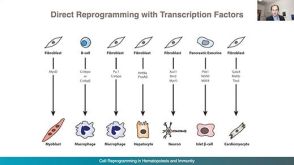
Reprogramming of T cells from human peripheral blood.
Vogt-Koyanagi-Harada (VKH) disease (and sympathetic ophthalmia) is an ocular inflammatory disease that is considered to be a cell-mediated autoimmune disease against melanocytes. The purpose of this study was to determine the Ags specific to VKH disease and to develop an animal model of VKH disease. We found that exposure of lymphocytes from patients with VKH disease to peptides (30-mer) derived from the tyrosinase family proteins led to significant proliferation of the lymphocytes. Immunization of these peptides into pigmented rats induced ocular and extraocular changes that highly resembled human VKH disease,and we suggest that an experimental VKH disease was induced in these rats. We conclude that VKH disease is an autoimmune disease against the tyrosinase family proteins.
View Publication

 EasySep™小鼠TIL(CD45)正选试剂盒


 沪公网安备31010102008431号