Keller GM (DEC 1995)
Current opinion in cell biology 7 6 862--9
In vitro differentiation of embryonic stem cells.
Under appropriate conditions in culture,embryonic stem cells will differentiate and form embryoid bodies that have been shown to contain cells of the hematopoietic,endothelial,muscle and neuronal lineages. Many aspects of the lineage-specific differentiation programs observed within the embryoid bodies reflect those found in the embryo,indicating that this model system provides access to early cell populations that develop in a normal fashion. Recent studies involving the differentiation of genetically altered embryonic stem cells highlight the potential of this in vitro differentiation system for defining the function of genes in early development.
View Publication
Chua SJ et al. (FEB 2009)
Biochemical and biophysical research communications 379 2 217--21
Neural progenitors, neurons and oligodendrocytes from human umbilical cord blood cells in a serum-free, feeder-free cell culture.
We have previously demonstrated that lineage negative cells (Lin(neg)) from umbilical cord blood (UCB) develop into multipotent cells capable of differentiation into bone,muscle,endothelial and neural cells. The objective of this study was to determine the optimal conditions required for Lin(neg) UCB cells to differentiate into neuronal cells and oligodendrocytes. We demonstrate that early neural stage markers (nestin,neurofilament,A2B5 and Sox2) are expressed in Lin(neg) cells cultured in FGF4,SCF,Flt3-ligand reprogramming culture media followed by the early macroglial cell marker O4. Early stage oligodendrocyte markers CNPase,GalC,Olig2 and the late-stage marker MOSP are observed,as is the Schwann cell marker PMP22. In summary,Lin(neg) UCB cells,when appropriately cultured,are able to exhibit characteristics of neuronal and macroglial cells that can specifically differentiate into oligodendrocytes and Schwann cells and express proteins associated with myelin production after in vitro differentiation.
View Publication
产品号#:
09600
09650
产品名:
StemSpan™ SFEM
StemSpan™ SFEM
Ray MK et al. (JUL 2016)
The Journal of biological chemistry jbc.M116.730853
CAT7 and cat7l long non-coding RNAs Tune Polycomb Repressive Complex 1 Function During Human and Zebrafish Development.
The essential functions of Polycomb Repressive Complex 1 (PRC1) in development and gene silencing are thought to involve long non-coding RNAs (lncRNAs),but few specific lncRNAs that guide PRC1 activity are known. We screened for lncRNAs which co-precipitate with PRC1 from chromatin and found candidates that impact Polycomb Group protein (PcG)-regulated gene expression in vivo. A novel lncRNA from this screen,CAT7,regulates expression and PcG binding at the MNX1 locus during early neuronal differentiation. CAT7 contains a unique tandem repeat domain which shares high sequence similarity to a non-syntenic zebrafish analog,cat7l. Defects caused by interference of cat7l RNA during zebrafish embryogenesis were rescued by human CAT7 RNA,enhanced by interference of a PRC1 component,and suppressed by interference of a known PRC1 target gene,demonstrating cat7l genetically interacts with a PRC1. We propose a model whereby PRC1 acts in concert with specific lncRNAs,and that CAT7/cat7l represent convergent lncRNAs that independently evolved to tune PRC1 repression at individual loci.
View Publication
Nguyen HX et al. (AUG 2014)
Journal of Comparative Neurology 522 12 2767--2783
Induction of early neural precursors and derivation of tripotent neural stem cells from human pluripotent stem cells under xeno-free conditions
Human embryonic stem cells (hESC) and induced pluripotent stem cells (hiPSC) can differentiate into many cell types and are important for regenerative medicine; however,further work is needed to reliably differentiate hESC and hiPSC into neural-restricted multipotent derivatives or specialized cell types under conditions that are free from animal products. Toward this goal,we tested the transition of hESC and hiPSC lines onto xeno-free (XF) / feeder-free conditions and evaluated XF substrate preference,pluripotency,and karyotype. Critically,XF transitioned H9 hESC,Shef4 hESC,and iPS6-9 retained pluripotency (Oct-4 and NANOG),proliferation (MKI67 and PCNA),and normal karyotype. Subsequently,XF transitioned hESC and hiPSC were induced with epidermal growth factor (EGF) and basic fibroblast growth factor (bFGF) to generate neuralized spheres containing primitive neural precursors,which could differentiate into astrocytes and neurons,but not oligoprogenitors. Further neuralization of spheres via LIF supplementation and attachment selection on CELLstart substrate generated adherent human neural stem cells (hNSC) with normal karyotype and high proliferation potential under XF conditions. Interestingly,adherent hNSC derived from H9,Shef4,and iPS6-9 differentiated into significant numbers of O4+ oligoprogenitors (∼20-30%) with robust proliferation; however,very few GalC+ cells were observed (∼2-4%),indicative of early oligodendrocytic lineage commitment. Overall,these data demonstrate the transition of multiple hESC and hiPSC lines onto XF substrate and media conditions,and a reproducible neuralization method that generated neural derivatives with multipotent cell fate potential and normal karyotype.
View Publication
产品号#:
05860
05880
05850
05857
05870
05875
85850
85857
85870
85875
产品名:
mTeSR™1
mTeSR™1
Pecho-Vrieseling E et al. (AUG 2014)
Nat Neurosci 17 8 1064--1072
Transneuronal propagation of mutant huntingtin contributes to non-cell autonomous pathology in neurons.
In Huntington's disease (HD),whether transneuronal spreading of mutant huntingtin (mHTT) occurs and its contribution to non-cell autonomous damage in brain networks is largely unknown. We found mHTT spreading in three different neural network models: human neurons integrated in the neural network of organotypic brain slices of HD mouse model,an ex vivo corticostriatal slice model and the corticostriatal pathway in vivo. Transneuronal propagation of mHTT was blocked by two different botulinum neurotoxins,each known for specifically inactivating a single critical component of the synaptic vesicle fusion machinery. Moreover,healthy human neurons in HD mouse model brain slices displayed non-cell autonomous changes in morphological integrity that were more pronounced when these neurons bore mHTT aggregates. Altogether,our findings suggest that transneuronal propagation of mHTT might be an important and underestimated contributor to the pathophysiology of HD.
View Publication
产品号#:
05850
05857
05870
05875
85850
85857
85870
85875
产品名:
mTeSR™1
mTeSR™1
Chen C et al. (JUL 2014)
Nature communications 5 4430
Role of astroglia in Down's syndrome revealed by patient-derived human-induced pluripotent stem cells.
Down's syndrome (DS),caused by trisomy of human chromosome 21,is the most common genetic cause of intellectual disability. Here we use induced pluripotent stem cells (iPSCs) derived from DS patients to identify a role for astrocytes in DS pathogenesis. DS astroglia exhibit higher levels of reactive oxygen species and lower levels of synaptogenic molecules. Astrocyte-conditioned medium collected from DS astroglia causes toxicity to neurons,and fails to promote neuronal ion channel maturation and synapse formation. Transplantation studies show that DS astroglia do not promote neurogenesis of endogenous neural stem cells in vivo. We also observed abnormal gene expression profiles from DS astroglia. Finally,we show that the FDA-approved antibiotic drug,minocycline,partially corrects the pathological phenotypes of DS astroglia by specifically modulating the expression of S100B,GFAP,inducible nitric oxide synthase,and thrombospondins 1 and 2 in DS astroglia. Our studies shed light on the pathogenesis and possible treatment of DS by targeting astrocytes with a clinically available drug.
View Publication
产品号#:
05850
05857
05870
05875
85850
85857
85870
85875
产品名:
mTeSR™1
mTeSR™1
Kim JJ et al. (DEC 2014)
Genomics data 2 10 139--143
Molecular effect of ethanol during neural differentiation of human embryonic stem cells in vitro.
Potential teratogenic effects of alcohol on fetal development have been documented. Especially studies have demonstrated deleterious effect of ethanol exposure on neuronal development in animal models and on the maintenance and differentiation of neuronal precursor cells derived from stem cells. To better understand the molecular effect of alcohol on the process of neural differentiation,we have performed gene expression microarray analysis on human embryonic stem cells being directed to neural rosettes and neural precursor cells in the presence of ethanol treatment. Here we provide detailed experimental methods,analysis and information associated with our data deposited into Gene Expression Omnibus (GEO) under GSE56906. Our data provide scientific insight on potential molecular effects of fetal alcohol exposure on neural differentiation of early embryo development.
View Publication

 EasySep™小鼠TIL(CD45)正选试剂盒


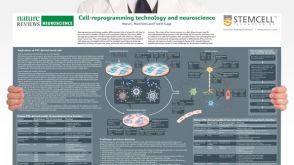 挂图Cell-Reprogramming Technology and Neuroscience Details on human iPSC-derived models of neuropsychiatric and neurodegenerative disorders

 沪公网安备31010102008431号