Okabe S et al. (SEP 1996)
Mechanisms of development 59 1 89--102
Development of neuronal precursor cells and functional postmitotic neurons from embryonic stem cells in vitro.
To understand the mechanism of the sequential restriction of multipotency of stem cells during development,we have established culture conditions that allow the differentiation of neuroepithelial precursor cells from embryonic stem (ES) cells. A highly enriched population of neuroepithelial precursor cells derived from ES cells proliferates in the presence of basic fibroblast growth factor (bFGF). These cells differentiate into both neurons and glia following withdrawal of bFGF. By further differentiating the cells in serum-containing medium,the neurons express a wide variety of neuron-specific genes and generate both excitatory and inhibitory synaptic connections. The expression pattern of position-specific neural markers suggests the presence of a variety of central nervous system (CNS) neuronal cell types. These findings indicate that neuronal precursor cells can be isolated from ES cells and that these cells can efficiently differentiate into functional post-mitotic neurons of diverse CNS structures.
View Publication
产品号#:
06902
06952
00321
00322
00323
00324
00325
产品名:
Cai S et al. (APR 2011)
Clinical cancer research : an official journal of the American Association for Cancer Research 17 8 2195--206
Humanized bone marrow mouse model as a preclinical tool to assess therapy-mediated hematotoxicity.
PURPOSE: Preclinical in vivo studies can help guide the selection of agents and regimens for clinical testing. However,one of the challenges in screening anticancer therapies is the assessment of off-target human toxicity. There is a need for in vivo models that can simulate efficacy and toxicities of promising therapeutic regimens. For example,hematopoietic cells of human origin are particularly sensitive to a variety of chemotherapeutic regimens,but in vivo models to assess potential toxicities have not been developed. In this study,a xenograft model containing humanized bone marrow is utilized as an in vivo assay to monitor hematotoxicity. EXPERIMENTAL DESIGN: A proof-of-concept,temozolomide-based regimen was developed that inhibits tumor xenograft growth. This regimen was selected for testing because it has been previously shown to cause myelosuppression in mice and humans. The dose-intensive regimen was administered to NOD.Cg-Prkdc(scid)IL2rg(tm1Wjl)/Sz (NOD/SCID/γchain(null)),reconstituted with human hematopoietic cells,and the impact of treatment on human hematopoiesis was evaluated. RESULTS: The dose-intensive regimen resulted in significant decreases in growth of human glioblastoma xenografts. When this regimen was administered to mice containing humanized bone marrow,flow cytometric analyses indicated that the human bone marrow cells were significantly more sensitive to treatment than the murine bone marrow cells and that the regimen was highly toxic to human-derived hematopoietic cells of all lineages (progenitor,lymphoid,and myeloid). CONCLUSIONS: The humanized bone marrow xenograft model described has the potential to be used as a platform for monitoring the impact of anticancer therapies on human hematopoiesis and could lead to subsequent refinement of therapies prior to clinical evaluation.
View Publication
产品号#:
03434
03444
04434
04444
84434
84444
产品名:
MethoCult™ GF M3434
MethoCult™ GF M3434
MethoCult™ H4434 Classic
MethoCult™ H4434 Classic
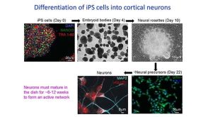
Wen Y and Jin S (OCT 2014)
Journal of Biotechnology 188 122--129
Production of neural stem cells from human pluripotent stem cells
Despite significant advances in commercially available media and kits and the differentiation approaches for human neural stem cell (NSC) generation,NSC production from the differentiation of human pluripotent stem cell (hPSC) is complicated by its time-consuming procedure,complex medium composition,and purification step. In this study,we developed a convenient and simplified NSC production protocol to meet the demand of NSC production. We demonstrated that NSCs can be generated efficiently without requirement of specific small molecules or embryoid body formation stage. Our experimental results suggest that a short suspension culture period may facilitate ectoderm lineage specification rather than endoderm or mesoderm lineage specification from hPSCs. The method developed in this study shortens the turnaround time of NSC production from both human embryonic stem cells (hESCs) and induced pluripotent stem cells (iPSCs) differentiation. It provides a straightforward and useful strategy for generating NSCs that can benefit a wide range of research applications for human brain research.
View Publication
产品号#:
05832
05850
05857
05870
05875
07923
85850
85857
85870
85875
产品名:
STEMdiff™ 神经花环选择试剂
Dispase (1 U/mL)
mTeSR™1
mTeSR™1
Fukuta M et al. (DEC 2014)
PLoS ONE 9 12 e112291
Derivation of mesenchymal stromal cells from pluripotent stem cells through a neural crest lineage using small molecule compounds with defined media
Neural crest cells (NCCs) are an embryonic migratory cell population with the ability to differentiate into a wide variety of cell types that contribute to the craniofacial skeleton,cornea,peripheral nervous system,and skin pigmentation. This ability suggests the promising role of NCCs as a source for cell-based therapy. Although several methods have been used to induce human NCCs (hNCCs) from human pluripotent stem cells (hPSCs),such as embryonic stem cells (ESCs) and induced pluripotent stem cells (iPSCs),further modifications are required to improve the robustness,efficacy,and simplicity of these methods. Chemically defined medium (CDM) was used as the basal medium in the induction and maintenance steps. By optimizing the culture conditions,the combination of the GSK3β inhibitor and TGFβ inhibitor with a minimum growth factor (insulin) very efficiently induced hNCCs (70-80%) from hPSCs. The induced hNCCs expressed cranial NCC-related genes and stably proliferated in CDM supplemented with EGF and FGF2 up to at least 10 passages without changes being observed in the major gene expression profiles. Differentiation properties were confirmed for peripheral neurons,glia,melanocytes,and corneal endothelial cells. In addition,cells with differentiation characteristics similar to multipotent mesenchymal stromal cells (MSCs) were induced from hNCCs using CDM specific for human MSCs. Our simple and robust induction protocol using small molecule compounds with defined media enabled the generation of hNCCs as an intermediate material producing terminally differentiated cells for cell-based innovative medicine.
View Publication
产品号#:
05850
05857
05870
05875
85850
85857
85870
85875
产品名:
mTeSR™1
mTeSR™1
Li Y et al. (MAR 2015)
PLoS ONE 10 3 e0118266
A comprehensive library of familial human amyotrophic lateral sclerosis induced pluripotent stem cells
Amyotrophic lateral sclerosis is a progressive disease characterized by the loss of upper and lower motor neurons,leading to paralysis of voluntary muscles. About 10% of all ALS cases are familial (fALS),among which 15-20% are linked to Cu/Zn superoxide dismutase (SOD1) mutations,usually inherited in an autosomal dominant manner. To date only one FDA approved drug is available which increases survival moderately. Our understanding of ALS disease mechanisms is largely derived from rodent model studies,however due to the differences between rodents and humans,it is necessary to have humanized models for studies of disease pathogenesis as well as drug development. Therefore,we generated a comprehensive library of a total 22 of fALS patient-specific induced pluripotent stem cell (iPSC) lines. These cells were thoroughly characterized before being deposited into the library. The library of cells includes a variety of C9orf72 mutations,sod1 mutations,FUS,ANG and FIG4 mutations. Certain mutations are represented with more than one line,which allows for studies of variable genetic backgrounds. In addition,these iPSCs can be successfully differentiated to astroglia,a cell type known to play a critical role in ALS disease progression. This library represents a comprehensive resource that can be used for ALS disease modeling and the development of novel therapeutics.
View Publication
产品号#:
05850
05857
05870
05875
07923
85850
85857
85870
85875
产品名:
Dispase (1 U/mL)
mTeSR™1
mTeSR™1
Carmona-Mora P et al. (OCT 2015)
Human Genetics 134 10 1099--1115
The nuclear localization pattern and interaction partners of GTF2IRD1 demonstrate a role in chromatin regulation
GTF2IRD1 is one of the three members of the GTF2I gene family,clustered on chromosome 7 within a 1.8 Mb region that is prone to duplications and deletions in humans. Hemizygous deletions cause Williams-Beuren syndrome (WBS) and duplications cause WBS duplication syndrome. These copy number variations disturb a variety of developmental systems and neurological functions. Human mapping data and analyses of knockout mice show that GTF2IRD1 and GTF2I underpin the craniofacial abnormalities,mental retardation,visuospatial deficits and hypersociability of WBS. However,the cellular role of the GTF2IRD1 protein is poorly understood due to its very low abundance and a paucity of reagents. Here,for the first time,we show that endogenous GTF2IRD1 has a punctate pattern in the nuclei of cultured human cell lines and neurons. To probe the functional relationships of GTF2IRD1 in an unbiased manner,yeast two-hybrid libraries were screened,isolating 38 novel interaction partners,which were validated in mammalian cell lines. These relationships illustrate GTF2IRD1 function,as the isolated partners are mostly involved in chromatin modification and transcriptional regulation,whilst others indicate an unexpected role in connection with the primary cilium. Mapping of the sites of protein interaction also indicates key features regarding the evolution of the GTF2IRD1 protein. These data provide a visual and molecular basis for GTF2IRD1 nuclear function that will lead to an understanding of its role in brain,behaviour and human disease.
View Publication
产品号#:
05850
05857
05870
05875
85850
85857
85870
85875
产品名:
mTeSR™1
mTeSR™1
Fuller HR et al. (JAN 2015)
Frontiers in cellular neuroscience 9 January 506
Spinal Muscular Atrophy Patient iPSC-Derived Motor Neurons Have Reduced Expression of Proteins Important in Neuronal Development.
Spinal muscular atrophy (SMA) is an inherited neuromuscular disease primarily characterized by degeneration of spinal motor neurons,and caused by reduced levels of the SMN protein. Previous studies to understand the proteomic consequences of reduced SMN have mostly utilized patient fibroblasts and animal models. We have derived human motor neurons from type I SMA and healthy controls by creating their induced pluripotent stem cells (iPSCs). Quantitative mass spectrometry of these cells revealed increased expression of 63 proteins in control motor neurons compared to respective fibroblasts,whereas 30 proteins were increased in SMA motor neurons vs. their fibroblasts. Notably,UBA1 was significantly decreased in SMA motor neurons,supporting evidence for ubiquitin pathway defects. Subcellular distribution of UBA1 was predominantly cytoplasmic in SMA motor neurons in contrast to nuclear in control motor neurons; suggestive of neurodevelopmental abnormalities. Many of the proteins that were decreased in SMA motor neurons,including beta III-tubulin and UCHL1,were associated with neurodevelopment and differentiation. These neuron-specific consequences of SMN depletion were not evident in fibroblasts,highlighting the importance of iPSC technology. The proteomic profiles identified here provide a useful resource to explore the molecular consequences of reduced SMN in motor neurons,and for the identification of novel biomarker and therapeutic targets for SMA.
View Publication

 EasySep™小鼠TIL(CD45)正选试剂盒


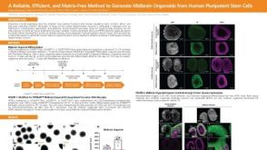 科学海报A Reliable, Efficient, and Matrix-Free Method to Generate Midbrain Organoids from Human Pluripotent Stem Cells

 沪公网安备31010102008431号