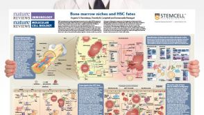
Bone Marrow Niches and HSC Fates
A detailed reference on signaling pathways in the bone marrow and how these influence HSC fate decisions; created in partnership with Nature Reviews Immunology and Nature Reviews Molecular Cell Biology
Zhao H et al. (JAN 2009)
Blood 113 3 505--16
The c-myb proto-oncogene and microRNA-15a comprise an active autoregulatory feedback loop in human hematopoietic cells.
The c-myb proto-oncogene encodes an obligate hematopoietic cell transcription factor important for lineage commitment,proliferation,and differentiation. Given its critical functions,c-Myb regulatory factors are of great interest but remain incompletely defined. Herein we show that c-Myb expression is subject to posttranscriptional regulation by microRNA (miRNA)-15a. Using a luciferase reporter assay,we found that miR-15a directly binds the 3'-UTR of c-myb mRNA. By transfecting K562 myeloid leukemia cells with a miR-15a mimic,functionality of binding was shown. The mimic decreased c-Myb expression,and blocked the cells in the G(1) phase of cell cycle. Exogenous expression of c-myb mRNA lacking the 3'-UTR partially rescued the miR-15a induced cell-cycle block. Of interest,the miR-15a promoter contained several potential c-Myb protein binding sites. Occupancy of one canonical c-Myb binding site was demonstrated by chromatin immunoprecipitation analysis and shown to be required for miR-15a expression in K562 cells. Finally,in studies using normal human CD34(+) cells,we showed that c-Myb and miR-15a expression were inversely correlated in cells undergoing erythroid differentiation,and that overexpression of miR-15a blocked both erythroid and myeloid colony formation in vitro. In aggregate,these findings suggest the presence of a c-Myb-miR-15a autoregulatory feedback loop of potential importance in human hematopoiesis.
View Publication
产品号#:
09500
产品名:
BIT 9500血清替代物
Singh KP et al. (JAN 2009)
Carcinogenesis 30 1 11--9
Treatment of mice with the Ah receptor agonist and human carcinogen dioxin results in altered numbers and function of hematopoietic stem cells.
The aryl hydrocarbon receptor (AhR) mediates the carcinogenicity of a family of environmental contaminants,the most potent being 2,3,7,8-tetrachlorodibenzo-p-dioxin (TCDD). Increased incidence of lymphoma and leukemia in humans is associated with TCDD exposure. Although AhR activation by TCDD has profound effects on the immune system,precise cellular and molecular mechanisms have yet to be determined. These studies tested the hypothesis that alteration of marrow populations following treatment of mice with TCDD is due to an effect on hematopoietic stem cells (HSCs). Treatment with TCDD resulted in an increased number and proliferation of bone marrow (BM) populations enriched for HSCs. There was a time-dependent decrease in B-lineage cells with a concomitant increase in myeloid populations. The decrease in the B-cell lineage colony-forming unit-preB progenitors along with a transient increase in myeloid progenitors were consistent with a skewing of lineage development from lymphoid to myeloid populations. However,HSCs from TCDD-treated mice exhibited diminished capacity to reconstitute and home to marrow of irradiated recipients. AhR messenger RNA was expressed in progenitor subsets but is downregulated during HSC proliferation. This result was consistent with the lack of response following the exposure of 5-fluorouracil-treated mice to TCDD. The direct exposure of cultured BM cells to TCDD inhibited the growth of immature hematopoietic progenitor cells,but not more mature lineage-restricted progenitors. Overall,these data are consistent with the hypothesis that TCDD,through AhR activation,alters the ability of HSCs to respond appropriately to signals within the marrow microenvironment.
View Publication
产品号#:
03231
产品名:
MethoCult™ M3231
Li X et al. (MAR 2009)
Human reproduction (Oxford,England) 24 3 580--9
ROCK inhibitor improves survival of cryopreserved serum/feeder-free single human embryonic stem cells.
BACKGROUND Efficient slow freezing protocols within serum-free and feeder-free culture systems are crucial for the clinical application of human embryonic stem (hES) cells. Frequently,however,hES cells must be cryopreserved as clumps when using conventional slow freezing protocols,leading to lower survival rates during freeze-thaw and limiting their recovery and growth efficiency after thawing,as well as limiting downstream applications that require single cell suspensions. We describe a novel method to increase freeze-thaw survival and proliferation rate of single hES cells in serum-free and feeder-free culture conditions. METHODS hES cells maintained on Matrigel-coated dishes were dissociated into single cells with Accutase and slow freezing. After thawing at 37 degrees C,cells were cultured in mTeSR medium supplemented with 10 microM of Rho-associated kinase inhibitor Y-27632 for 1 day. RESULTS The use of Y-27632 and Accutase significantly increases the survival of single hES cells after thawing compared with a control group (P textless 0.01). Furthermore,by treatment of hES cell aggregates with EGTA to disrupt cell-cell interaction,we show that Y-27632 treatment does not directly affect hES cell apoptosis. Even in the presence of Y-27632,hES cells deficient in cell-cell interaction undergo apoptosis. Y-27632-treated freeze-thawed hES cells retain typical morphology,stable karyotype,expression of pluripotency markers and the potential to differentiate into derivatives of all three germ layers after long-term culture. CONCLUSIONS The method described here allows for cryopreservation of single hES cells in serum-free and feeder-free conditions and therefore we believe this method will be ideal for current and future hES cell applications that are targeted towards a therapeutic end-point.
View Publication
产品号#:
05850
05857
05870
05875
72302
72304
72307
72308
85850
85857
85870
85875
100-1044
产品名:
Y-27632(二盐酸盐)
Y-27632(二盐酸盐)
Y-27632(二盐酸盐)
Y-27632(二盐酸盐)
mTeSR™1
mTeSR™1
Y-27632(二盐酸盐)
Li Z et al. (FEB 2009)
Journal of cellular biochemistry 106 2 194--9
Transplantation of human embryonic stem cell-derived endothelial cells for vascular diseases.
Using endothelial cells for therapeutic angiogenesis/vasculogenesis of ischemia diseases has led to exploring human embryonic stem cells (hESCs) as a potentially unlimited source for endothelial progenitor cells. With their capacity for self-renewal and pluripotency,hESCs and their derived endothelial cells (hESC-ECs) may be more advantageous than other endothelial cells obtained from diseased populations. However,hESC-ECs' poor differentiation efficiency and poorly characterized in vivo function after transplantation present significant challenges for their future clinical application. This review will focus on the differentiation pathways of hESCs and their therapeutic potential for vascular diseases,as well as the monitoring of transplanted cells' fate via molecular imaging. Finally,cell enhancement strategies to improve the engraftment efficiency of hESC-ECs will be discussed.
View Publication
产品号#:
05850
05857
05870
05875
85850
85857
85870
85875
产品名:
mTeSR™1
mTeSR™1
Praetor A et al. (FEB 2009)
Blood 113 9 1919--28
Genetic deletion of JAM-C reveals a role in myeloid progenitor generation.
Hematopoietic stem cells (HSCs) have the capacity to self-renew and continuously differentiate into all blood cell lineages throughout life. At each branching point during differentiation,interactions with the environment are key in the generation of daughter cells with distinct fates. Here,we examined the role of the cell adhesion molecule JAM-C,a protein known to mediate cellular polarity during spermatogenesis,in hematopoiesis. We show that murine JAM-C is highly expressed on HSCs in the bone marrow (BM). Expression correlates with self-renewal,the highest being on long-term repopulating HSCs,and decreases with differentiation,which is maintained longest among myeloid committed progenitors. Inclusion of JAM-C as a sole marker on lineage-negative BM cells yields HSC enrichments and long-term multilineage reconstitution when transferred to lethally irradiated mice. Analysis of Jam-C-deficient mice showed that two-thirds die within 48 hours after birth. In the surviving animals,loss of Jam-C leads to an increase in myeloid progenitors and granulocytes in the BM. Stem cells and myeloid cells from fetal liver are normal in number and homing to the BM. These results provide evidence that JAM-C defines HSCs in the BM and that JAM-C plays a role in controlling myeloid progenitor generation in the BM.
View Publication
产品号#:
03434
03444
产品名:
MethoCult™ GF M3434
MethoCult™ GF M3434
Lin H et al. (MAR 2009)
Experimental biology and medicine (Maywood,N.J.) 234 3 342--53
Maitake beta-glucan enhances umbilical cord blood stem cell transplantation in the NOD/SCID mouse.
Beta glucans are cell wall constituents of yeast,fungi and bacteria,as well as mushrooms and barley. Glucans are not expressed on mammalian cells and are recognized as pathogen-associated molecular patterns (PAMPS) by pattern recognition receptors (PRR). Beta glucans have potential activity as biological response modifiers for hematopoiesis and enhancement of bone marrow recovery after injury. We have reported that Maitake beta glucan (MBG) enhanced mouse bone marrow (BMC) and human umbilical cord blood (CB) cell granulocyte-monocyte colony forming unit (GM-CFU) activity in vitro and protected GM-CFU forming stem cells from doxorubicin (DOX) toxicity. The objective of this study was to determine the effects of MBG on expansion of phenotypically distinct subpopulations of progenitor and stem cells in CB from full-term infants cultured ex vivo and on homing and engraftment in vivo in the nonobese diabetic/severe combined immunodeficient (NOD/SCID) mouse. MBG promoted a greater expansion of CD34+CD33+CD38- human committed hematopoietic progenitor (HPC) cells compared to the conventional stem cell culture medium (P = 0.002 by ANOVA). CD34+CXCR4+CD38- early,uncommitted human hematopoietic stem cell (HSC) numbers showed a trend towards increase in response to MBG. The fate of CD34+ enriched CB cells after injection into the sublethally irradiated NOS/SCID mouse was evaluated after retrieval of xenografted human CB from marrow and spleen by flow cytometric analysis. Oral administration of MBG to recipient NOS/SCID mice led to enhanced homing at 3 days and engraftment at 6 days in mouse bone marrow (P = 0.002 and P = 0.0005,respectively) compared to control mice. More CD34+ human CB cells were also retrieved from mouse spleen in MBG treated mice at 6 days after transplantation. The studies suggest that MBG promotes hematopoiesis through effects on CD34+ progenitor cell expansion ex vivo and when given to the transplant recipient could enhance CD34+ precursor cell homing and support engraftment.
View Publication
产品号#:
02690
09600
09650
09850
15026
15066
产品名:
StemSpan™ CC100
StemSpan™ SFEM
StemSpan™ SFEM
RosetteSep™人造血祖细胞富集抗体混合物
RosetteSep™人造血祖细胞富集抗体混合物
Mellick AS et al. (SEP 2010)
Cancer research 70 18 7273--82
Using the transcription factor inhibitor of DNA binding 1 to selectively target endothelial progenitor cells offers novel strategies to inhibit tumor angiogenesis and growth.
Tumor angiogenesis is essential for malignant growth and metastasis. Bone marrow (BM)-derived endothelial progenitor cells (EPC) contribute to angiogenesis-mediated tumor growth. EPC ablation can reduce tumor growth; however,the lack of a marker that can track EPCs from the BM to tumor neovasculature has impeded progress in understanding the molecular mechanisms underlying EPC biology. Here,we report the use of transgenic mouse and lentiviral models to monitor the BM-derived compartment of the tumor stroma; this approach exploits the selectivity of the transcription factor inhibitor of DNA binding 1 (Id1) for EPCs to track EPCs in the BM,blood,and tumor stroma,as well as mature EPCs. Acute ablation of BM-derived EPCs using Id1-directed delivery of a suicide gene reduced circulating EPCs and yielded significant defects in angiogenesis-mediated tumor growth. Additionally,use of the Id1 proximal promoter to express microRNA-30-based short hairpin RNA inhibited the expression of critical EPC-intrinsic factors,confirming that signaling through vascular endothelial growth factor receptor 2 is required for EPC-mediated tumor biology. By exploiting the selectivity of Id1 gene expression in EPCs,our results establish a strategy to track and target EPCs in vivo,clarifying the significant role that EPCs play in BM-mediated tumor angiogenesis.
View Publication
产品号#:
09600
09650
产品名:
StemSpan™ SFEM
StemSpan™ SFEM
Gallego MJ et al. (JAN 2010)
Stem cell research & therapy 1 4 28
The pregnancy hormones human chorionic gonadotropin and progesterone induce human embryonic stem cell proliferation and differentiation into neuroectodermal rosettes.
INTRODUCTION: The physiological signals that direct the division and differentiation of the zygote to form a blastocyst,and subsequent embryonic stem cell division and differentiation during early embryogenesis,are unknown. Although a number of growth factors,including the pregnancy-associated hormone human chorionic gonadotropin (hCG) are secreted by trophoblasts that lie adjacent to the embryoblast in the blastocyst,it is not known whether these growth factors directly signal human embryonic stem cells (hESCs).backslashnbackslashnMETHODS: Here we used hESCs as a model of inner cell mass differentiation to examine the hormonal requirements for the formation of embryoid bodies (EB's; akin to blastulation) and neuroectodermal rosettes (akin to neurulation).backslashnbackslashnRESULTS: We found that hCG promotes the division of hESCs and their differentiation into EB's and neuroectodermal rosettes. Inhibition of luteinizing hormone/chorionic gonadotropin receptor (LHCGR) signaling suppresses hESC proliferation,an effect that is reversed by treatment with hCG. hCG treatment rapidly upregulates steroidogenic acute regulatory protein (StAR)-mediated cholesterol transport and the synthesis of progesterone (P4). hESCs express P4 receptor A,and treatment of hESC colonies with P4 induces neurulation,as demonstrated by the expression of nestin and the formation of columnar neuroectodermal cells that organize into neural tubelike rosettes. Suppression of P4 signaling by withdrawing P4 or treating with the P4-receptor antagonist RU-486 inhibits the differentiation of hESC colonies into EB's and rosettes.backslashnbackslashnCONCLUSIONS: Our findings indicate that hCG signaling via LHCGR on hESC promotes proliferation and differentiation during blastulation and neurulation. These findings suggest that trophoblastic hCG secretion and signaling to the adjacent embryoblast could be the commencement of trophic support by placental tissues in the growth and development of the human embryo.
View Publication
产品号#:
05850
05857
05870
05875
85850
85857
85870
85875
产品名:
mTeSR™1
mTeSR™1
Cantu' C et al. (JAN 2011)
Nucleic acids research 39 2 486--501
A highly conserved SOX6 double binding site mediates SOX6 gene downregulation in erythroid cells.
The Sox6 transcription factor plays critical roles in various cell types,including erythroid cells. Sox6-deficient mice are anemic due to impaired red cell maturation and show inappropriate globin gene expression in definitive erythrocytes. To identify new Sox6 target genes in erythroid cells,we used the known repressive double Sox6 consensus within the εy-globin promoter to perform a bioinformatic genome-wide search for similar,evolutionarily conserved motifs located within genes whose expression changes during erythropoiesis. We found a highly conserved Sox6 consensus within the Sox6 human gene promoter itself. This sequence is bound by Sox6 in vitro and in vivo,and mediates transcriptional repression in transient transfections in human erythroleukemic K562 cells and in primary erythroblasts. The binding of a lentiviral transduced Sox6FLAG protein to the endogenous Sox6 promoter is accompanied,in erythroid cells,by strong downregulation of the endogenous Sox6 transcript and by decreased in vivo chromatin accessibility of this region to the PstI restriction enzyme. These observations suggest that the negative Sox6 autoregulation,mediated by the double Sox6 binding site within its own promoter,may be relevant to control the Sox6 transcriptional downregulation that we observe in human erythroid cultures and in mouse bone marrow cells in late erythroid maturation.
View Publication
Yang J et al. (DEC 2010)
Journal of Biological Chemistry 285 51 40303--11
Induced pluripotent stem cells can be used to model the genomic imprinting disorder Prader-Willi syndrome.
The recent discovery of induced pluripotent stem cell (iPSC) technology provides an invaluable tool for creating in vitro representations of human genetic conditions. This is particularly relevant for those diseases that lack adequate animal models or where the species comparison is difficult,e.g. imprinting diseases such as the neurogenetic disorder Prader-Willi syndrome (PWS). However,recent reports have unveiled transcriptional and functional differences between iPSCs and embryonic stem cells that in cases are attributable to imprinting errors. This has suggested that human iPSCs may not be useful to model genetic imprinting diseases. Here,we describe the generation of iPSCs from a patient with PWS bearing a partial translocation of the paternally expressed chromosome 15q11-q13 region to chromosome 4. The resulting iPSCs match all standard criteria of bona fide reprogramming and could be readily differentiated into tissues derived from the three germ layers,including neurons. Moreover,these iPSCs retain a high level of DNA methylation in the imprinting center of the maternal allele and show concomitant reduced expression of the disease-associated small nucleolar RNA HBII-85/SNORD116. These results indicate that iPSCs may be a useful tool to study PWS and perhaps other genetic imprinting diseases as well.
View Publication

 EasySep™小鼠TIL(CD45)正选试剂盒


 挂图Bone Marrow Niches and HSC Fates A detailed reference on signaling pathways in the bone marrow and how these influence HSC fate decisions; created in partnership with Nature Reviews Immunology and Nature Reviews Molecular Cell Biology

 沪公网安备31010102008431号