
 EasySep™小鼠TIL(CD45)正选试剂盒


产品号 #08610_C
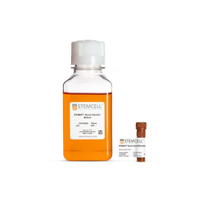
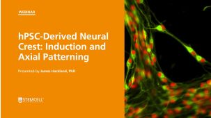
用于建立 hPSC 衍生神经嵴细胞的细胞培养试剂盒
若您需要咨询产品或有任何技术问题,请通过官方电话 400 885 9050 或邮箱 info.cn@stemcell.com 与我们联系。
用于建立 hPSC 衍生神经嵴细胞的细胞培养试剂盒
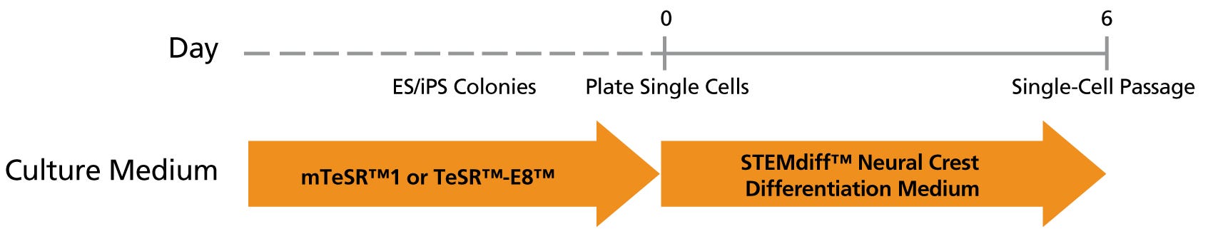
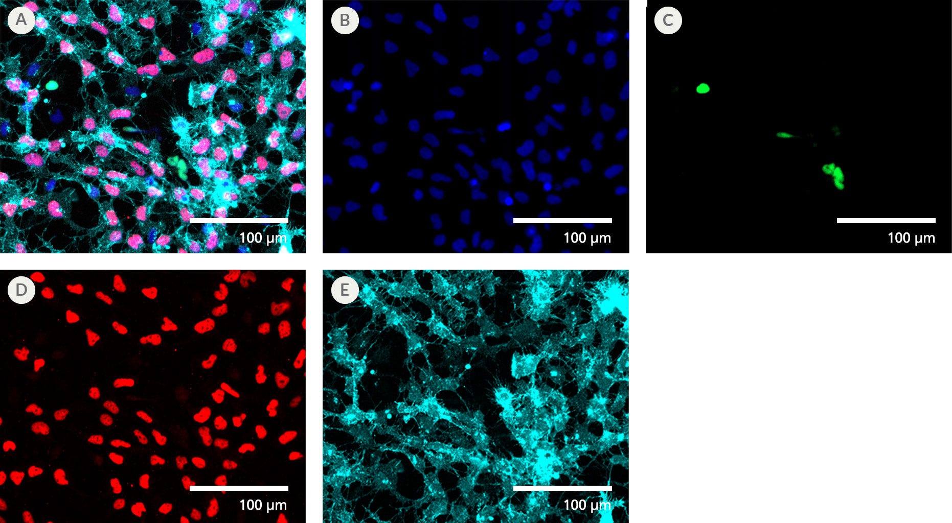
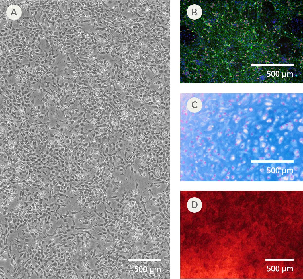
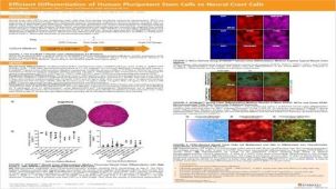
STEMdiff™ 神经嵴分化试剂盒提供无血清培养基,使人胚胎干细胞(ES细胞)和诱导多能干细胞(iPS细胞)分化为神经嵴细胞。这些神经嵴细胞以 SOX10 和 CD271 等神经嵴标志物为特征,并可进一步分化为多种下游细胞类型,包括软骨细胞、成骨细胞和周围神经元。
该培养基兼容在以下培养体系中维持的人ES和iPS细胞:mTeSR™1(目录编号:85850)、mTeSR™ Plus(目录编号:100-0276)或 TeSR™-E8™(目录编号:05990)。
分类
专用培养基
细胞类型
神经细胞,PSC衍生,神经干/祖细胞,神经元,多能干细胞
种属
人
应用
细胞培养,分化,细胞毒性检测
品牌
STEMdiff
研究领域
疾病建模,药物发现和毒理检测,神经科学,干细胞生物学
制剂类别
无血清
请在《产品说明书》中查找相关支持信息和使用说明,或浏览下方更多实验方案。
本产品专为以下研究领域设计,适用于工作流程中的高亮阶段。探索这些工作流程,了解更多我们为各研究领域提供的其他配套产品。
| 物种 | 人 |
|---|---|
| 配方 | 无血清 |
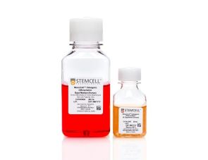
用于体外诱导人MSC分化为成骨细胞

无血清神经添加物(50X)
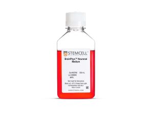
提升神经元功能的无血清基础培养基
<p>采用磁珠解离技术,对来源于人多能干细胞分化的神经嵴细胞中的 CD271⁺ 细胞进行免疫磁珠正选分离</p>
一次性使用、免维持的人诱导多能干细胞,冷冻
在线联系


 沪公网安备31010102008431号