
 EasySep™小鼠TIL(CD45)正选试剂盒


产品号 #05010_C
用于将人 PSC 分化为心室肌细胞以及长期维持人PSC衍生心肌细胞的无血清培养基
若您需要咨询产品或有任何技术问题,请通过官方电话 400 885 9050 或邮箱 info.cn@stemcell.com 与我们联系。
用于将人 PSC 分化为心室肌细胞以及长期维持人PSC衍生心肌细胞的无血清培养基
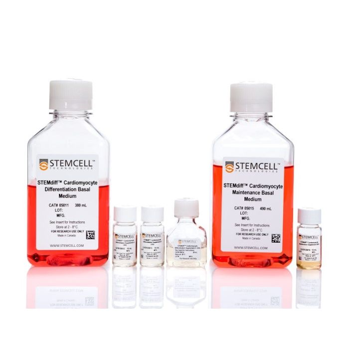
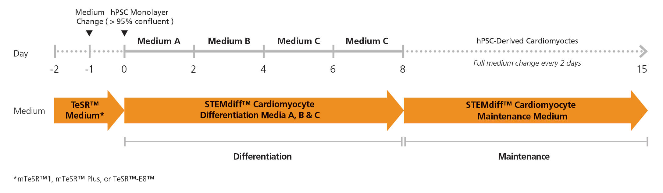
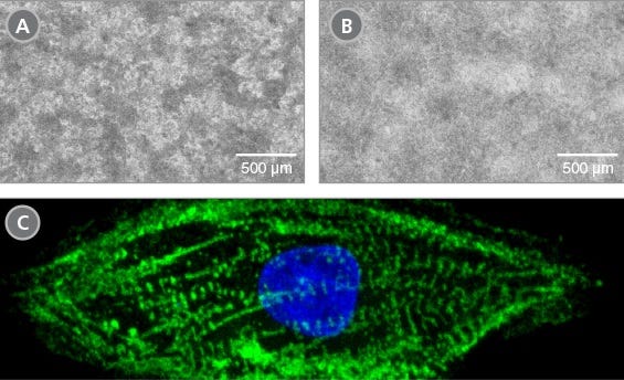
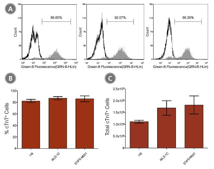
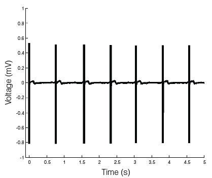
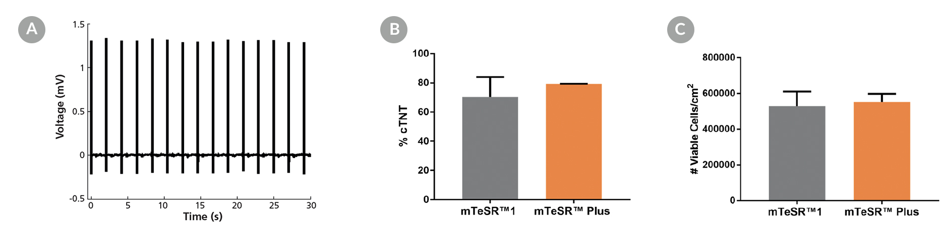
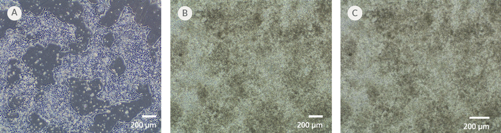
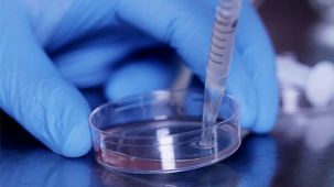
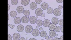
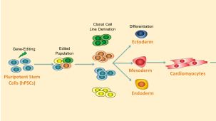
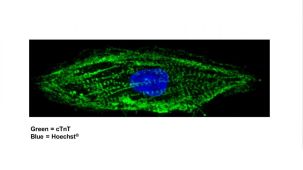
STEMdiff™ 心室肌细胞分化试剂盒(产品号 #05010)包含用于将人胚胎干细胞(ES)和诱导多能干细胞(iPS)(统称为人多能干细胞 [hPSCs])分化为心室肌细胞(肌钙蛋白T阳性 [cTnT+])的培养基,以及用于维持 hPSC 衍生心肌细胞的培养基。该试剂盒为无血清体系,可用于从以mTeSR™1(产品号 #85850)、mTeSR™ Plus(产品号 #100-0276)、TeSR™-AOF(产品号 #100-0401)或TeSR™-E8™(产品号 #05990)培养的hPSC团块中诱导分化出心室肌细胞。这些细胞中超过80%为cTnT+。平均每12孔板的单孔可收获1 x 10^6个细胞。
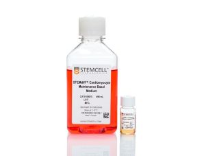
STEMdiff™ 心肌细胞维持试剂盒(产品号 #05020)包含维持基础培养基和补充剂,可用于长期维持hPSC衍生的心肌细胞一个月或更长时间。这些心肌细胞可用于各种下游应用和分析。
注:本产品原名为“STEMdiff™ 心肌细胞分化试剂盒”;产品本身和生产流程未发生变化,但名称已更新,以更准确地反映生成的细胞类型。
分类
专用培养基
细胞类型
心肌细胞,PSC衍生
种属
人
应用
细胞培养,分化,培养
品牌
STEMdiff
研究领域
疾病建模,药物发现和毒理检测,干细胞生物学
制剂类别
无血清
请在《产品说明书》中查找相关支持信息和使用说明,或浏览下方更多实验方案。
本产品专为以下研究领域设计,适用于工作流程中的高亮阶段。探索这些工作流程,了解更多我们为各研究领域提供的其他配套产品。
| 物种 | 人 |
|---|---|
| 配方 | 无血清 |
<p>用于长期维持培养人 PSC 衍生心肌细胞的培养基</p>
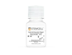
用于解离hPSC衍生的心肌细胞
用于复苏和培养 hPSC 衍生心肌细胞的培养基
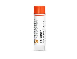
一次性使用、免维持的人诱导多能干细胞,冷冻
质量保证:
本产品仅供科研使用,除非另有说明,不得用于人体或动物的诊断或治疗用途。如需了解 STEMCELL 的质量体系,请访问www.stemcell.cn/compliance
在线联系


 沪公网安备31010102008431号