
 EasySep™小鼠TIL(CD45)正选试剂盒


产品号 #05455_C
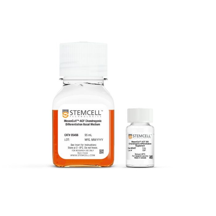
用于MSCs向软骨细胞分化的无动物成分培养基
若您需要咨询产品或有任何技术问题,请通过官方电话 400 885 9050 或邮箱 info.cn@stemcell.com 与我们联系。
用于MSCs向软骨细胞分化的无动物成分培养基
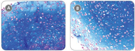
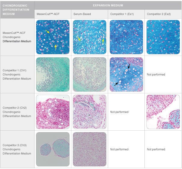
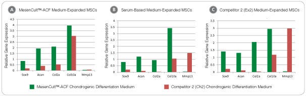
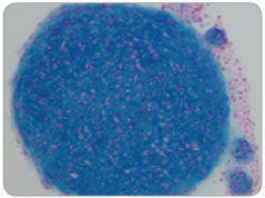
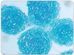
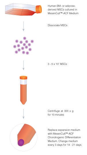
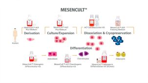
使用MesenCult™-ACF软骨分化试剂盒,从人间充质基质细胞(MSCs)和hPSC衍生的间充质祖细胞中生成软骨谱系细胞(包括软骨细胞)。这种完全无动物成分(ACF)的配方可从低至3×10⁵个细胞开始实现高效的软骨生成分化和基质沉积。生成的软骨细胞最早在第14天即可检测到软骨生成。该培养基与源自骨髓、脂肪组织或滑膜的MSC兼容,并适用于在含血清或ACF条件下扩增的细胞。
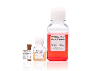
将此试剂盒作为完整的MSC功能评估工作流程的一部分使用。与MesenCult™脂肪生成分化试剂盒(人)和MesenCult™成骨分化试剂盒(人)配合使用,以评估三谱系分化潜能并验证MSC的身份和功能。这些即用型试剂能够可靠地评估MSC的身份和功能,同时确保在整个MesenCult™工作流程中获得可重复的谱系特异性结果。
分类
专用培养基
细胞类型
软骨细胞,间充质干/祖细胞
种属
人
应用
细胞培养,分化
品牌
MesenCult
研究领域
药物发现和毒性检测,干细胞生物学
制剂类别
不含动物成分,无血清
请在《产品说明书》中查找相关支持信息和使用说明,或浏览下方更多实验方案。
本产品专为以下研究领域设计,适用于工作流程中的高亮阶段。探索这些工作流程,了解更多我们为各研究领域提供的其他配套产品。
| 物种 | 人 |
|---|---|
| 配方 | 不含动物成分, 无血清 |
人MSCs向脂肪细胞分化的培养基
人干细胞和祖细胞解离试剂盒
在线联系

 沪公网安备31010102008431号