
 EasySep™小鼠TIL(CD45)正选试剂盒


产品号 #01700_C
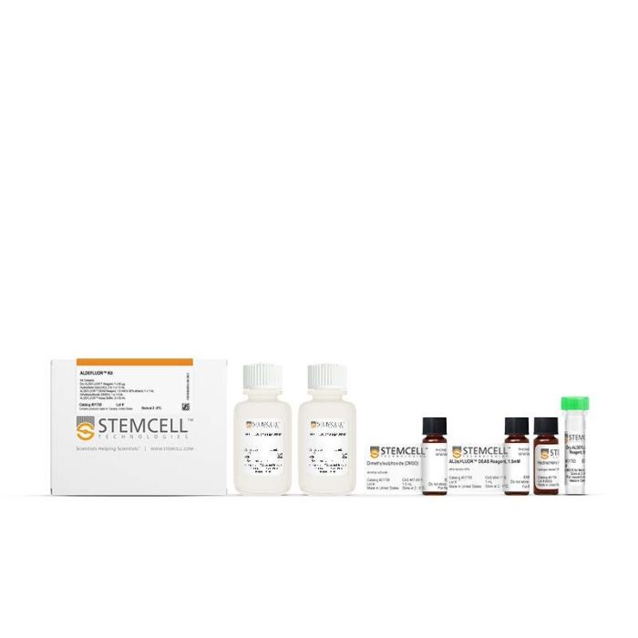
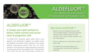
用于鉴定、评估和分离表达高水平ALDH的干细胞和祖细胞
若您需要咨询产品或有任何技术问题,请通过官方电话 400 885 9050 或邮箱 info.cn@stemcell.com 与我们联系。
用于鉴定、评估和分离表达高水平ALDH的干细胞和祖细胞
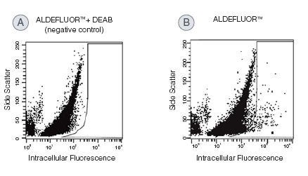
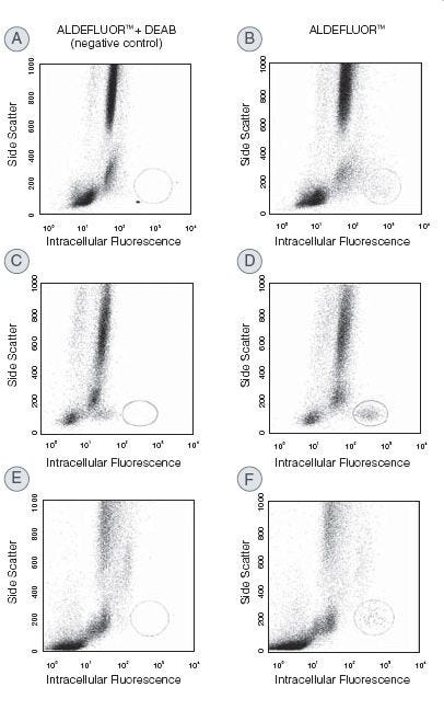
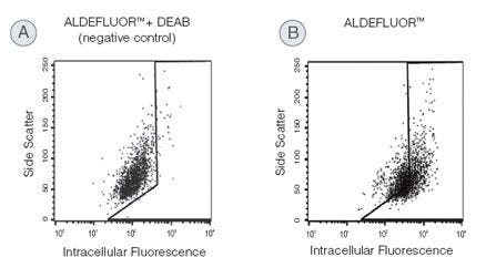
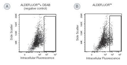
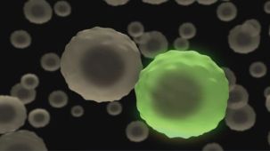
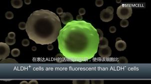
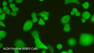
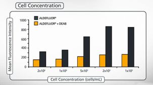
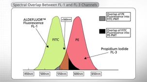
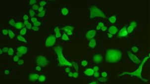
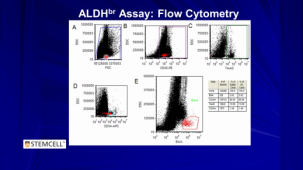
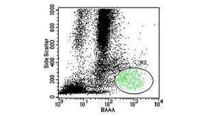
用ALDEFLUOR™试剂盒鉴定和分离表达醛脱氢酶(ALDH)的活细胞。与传统方法相比,该检测方法不需要抗体染色。已有研究报道,不同谱系的正常和癌症前体细胞中普遍存在高水平 ALDH 表达。ALDEFLUOR™检测是一种被广泛发表的用于检测ALDH 高表达(ALDHbr)细胞的非免疫学方法,可用于检测造血、乳腺、内皮、间充质、神经和其他组织中的癌细胞。试剂盒中包含的ALDEFLUOR™DEAB试剂和ALDEFLUOR™ 检测缓冲液支持检测的最佳效果,二者也可单独购买。该试剂盒与标准流式细胞仪兼容,可用于 ALDHbr 细胞的后续分析,并与标准细胞分选仪兼容,可用于进一步纯化和表征。查看我们的其他资源,以了解更多关于ALDEFLUOR™试剂系统的信息。
细胞类型
脑肿瘤干细胞,癌细胞及细胞系,造血干/祖细胞,乳腺细胞,间充质干/祖细胞,神经干/祖细胞,其他物种
种属
人,小鼠,非人灵长类,其他物种,大鼠
应用
流式细胞术
品牌
ALDEFLUOR
研究领域
癌症,上皮细胞研究,神经科学,干细胞生物学
CAS 编号
7647-01-0
请在《产品说明书》中查找相关支持信息和使用说明,或浏览下方更多实验方案。
本产品专为以下研究领域设计,适用于工作流程中的高亮阶段。探索这些工作流程,了解更多我们为各研究领域提供的其他配套产品。
| 物种 | 人, 其它物种, 大鼠, 小鼠, 非人灵长类 |
|---|---|
| Cas Number | 7647-01-0 |

用于ALDEFLUOR™试剂盒的附加检测缓冲液
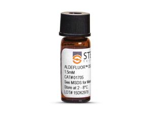
在ALDEFLUOR™检测系统中用作对照的ALDH酶抑制剂
用于人乳腺球和肿瘤球的培养
小鼠Monoclonal IgG1抗体,抗人CD34
在线联系


 沪公网安备31010102008431号