
 EasySep™小鼠TIL(CD45)正选试剂盒


产品号 #05620
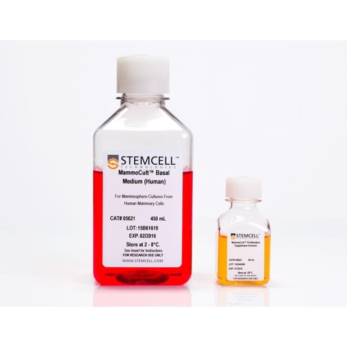
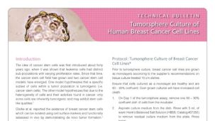
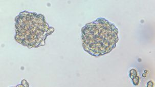
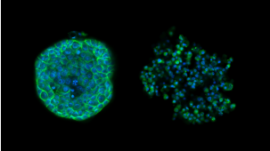
用于人乳腺球和肿瘤球的培养
若您需要咨询产品或有任何技术问题,请通过官方电话 400 885 9050 或邮箱 info.cn@stemcell.com 与我们联系。
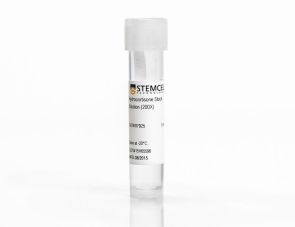
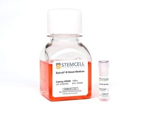
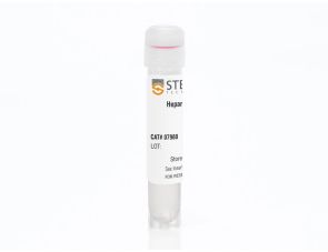
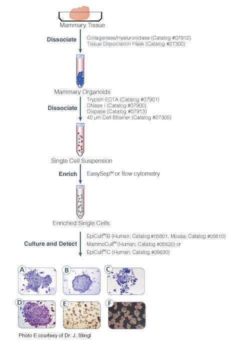
MammoCult™培养基(人源)是一种无血清、不含雌激素和孕酮的培养基,专为培养正常人原代乳腺组织来源的乳腺球和人乳腺癌细胞系来源的肿瘤球而优化。配制完整的MammoCult™培养基还需使用氢化可的松储备液(产品号#07925)和肝素溶液(产品号#07980)。
分类
专用培养基
细胞类型
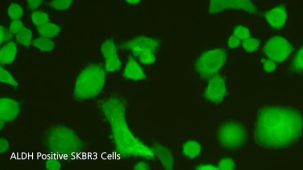
癌细胞及细胞系,乳腺细胞
种属
人
应用
细胞培养,培养,球状体培养
品牌
MammoCult
研究领域
癌症,上皮细胞研究
制剂类别
无血清
请在《产品说明书》中查找相关支持信息和使用说明,或浏览下方更多实验方案。
本产品专为以下研究领域设计,适用于工作流程中的高亮阶段。探索这些工作流程,了解更多我们为各研究领域提供的其他配套产品。
| 物种 | 人 |
|---|---|
| 配方 | 无血清 |
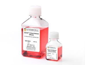
用于小鼠乳腺上皮细胞的培养和评估
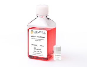
用于培养人乳腺上皮细胞
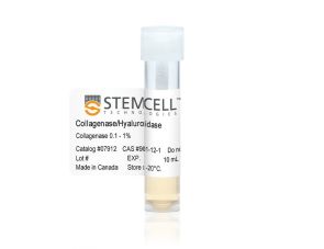
配置于DMEM中的10 X 胶原酶/透明质酸酶
细胞培养补充剂
在线联系


 沪公网安备31010102008431号