
 EasySep™小鼠TIL(CD45)正选试剂盒


产品号 #(选择产品)
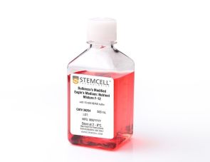
用于人肠道类器官扩增与分化的优化培养基
若您需要咨询产品或有任何技术问题,请通过官方电话 400 885 9050 或邮箱 info.cn@stemcell.com 与我们联系。
无血清且无条件培养基的人肠道类器官扩增与分化培养基
用于人肠道类器官扩增与分化的优化培养基
使用 IntestiCult™ Plus 类器官生长培养基(IntestiCult™ Plus),可增强肠道类器官的生理相关性,更好地反映人体内肠道的复杂性。与传统扩增和分化阶段分开进行的培养流程不同,IntestiCult™ Plus 是一种完整的、无血清、无条件培养基成分的配方,旨在简化肠道类器官培养流程,扩增与分化同步进行。
使用 IntestiCult™ Plus 培养的类器官可形成具有区域特异性的芽状和隐窝样结构,与第一代系统(如 IntestiCult™ Organoid Growth Medium(人;产品号 #06010)和 IntestiCult™ Organoid Differentiation Medium(人;产品号 #100-0214))生成的类器官相比,能更准确地再现肠上皮结构。IntestiCult™ Plus 在维持 Lgr5+ 干细胞高效扩增的同时,促进关键肠道细胞类型(如潘氏细胞、肠内分泌细胞、嗜铬细胞、杯状细胞和簇状细胞)的稳定生成。
该模型还可适用于生成无 Wnt 培养基,用于小肠分化等研究,或用于培养 Wnt 非依赖性的结直肠癌类器官。通过维持生理平衡的细胞类型组成,无论是进行疾病建模、药物评估还是毒性检测,IntestiCult™ Plus都能让您获得更可靠的实验结果。
若您计划将此产品用于商业目的,请联系 HUB Organoids B.V.(www.huborganoids.nl)以获取商业使用许可或了解相关授权信息。
请在《产品说明书》中查找相关支持信息和使用说明,或浏览下方更多实验方案。
Legal Statement:
This product was developed under a license to intellectual property owned by Hubrecht Organoid Technology (HUB). This product is sold for research use only. Purchase of this product does not include the right to use it for drug screening aiming for commercial gain, equipment validation, biobanking, or for other commercial purposes. Purchasers wishing to use the product for purposes other than basic research use should contact HUB at www.huborganoids.nl to obtain a further license. Purchasers may apply for a License from HUB, which will not be unreasonably withheld by HUB.
质量保证:
本产品仅供科研使用,除非另有说明,不得用于人体或动物的诊断或治疗用途。如需了解 STEMCELL 的质量体系,请访问 www.stemcell.cn/compliance
在线联系


 沪公网安备31010102008431号