
 EasySep™小鼠TIL(CD45)正选试剂盒


产品号 #100-1079_C
磁珠法总核酸(DNA和RNA)提取试剂盒
若您需要咨询产品或有任何技术问题,请通过官方电话 400 885 9050 或邮箱 info.cn@stemcell.com 与我们联系。
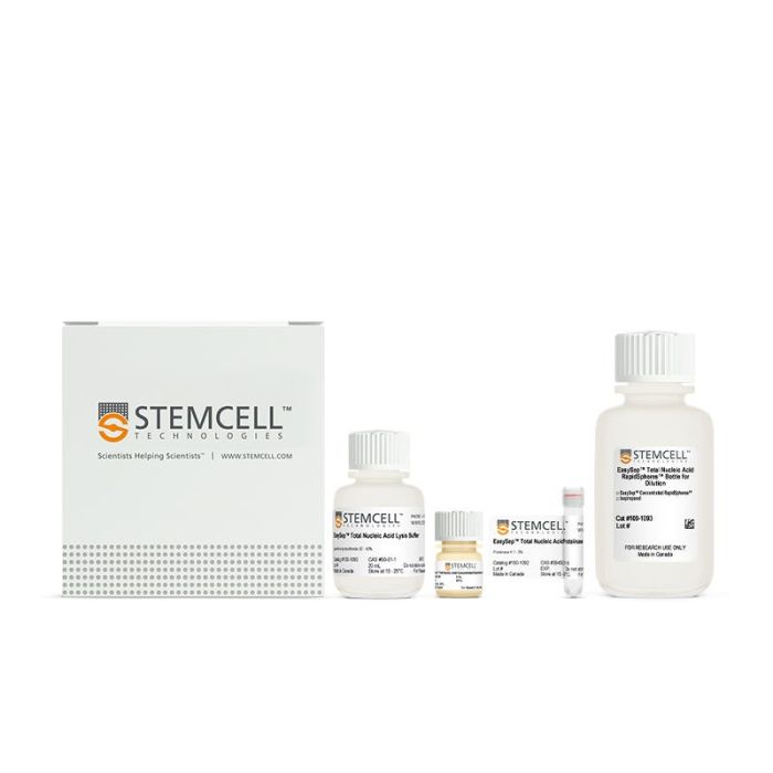
磁珠法总核酸(DNA和RNA)提取试剂盒
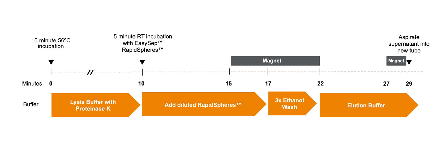
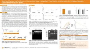
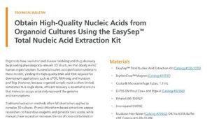
高效地从全血、细胞悬液(如 PBMCs、hPSCs、培养细胞)及 使用EasySep™ 分选的细胞中高效地提取总核酸(DNA 和 RNA)或仅提取 RNA。EasySep™ 总核酸提取试剂盒采用磁珠技术,配合简便且可扩展的操作流程,无需使用离心柱或有毒试剂。使用该试剂盒提取的核酸纯度高,可直接用于如qPCR等下游应用。
使用本试剂盒,可通过 EasySep™ 总核酸 RapidSpheres™ 磁珠对样本中的总核酸或 RNA 进行磁珠标记。随后无需分离柱,只需磁极(需单独购买)处理样本。去除未标记的杂质成分后,从磁极上取下样本并回收被磁珠标记的核酸。
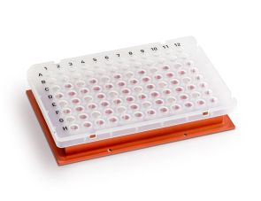
该试剂盒提供了两种方案:使用 1.7 mL 微量离心管配合 ErythroClear™ 磁极(产品号 #01737)进行总核酸或 RNA 提取;或在 96 孔 PCR 板中配合96孔PCR磁力板(产品号 #100-1304)实现高通量提取。
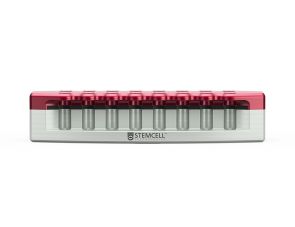
磁极兼容性
ErythroClear™磁极(产品号 #01737)
96孔PCR微孔板磁极(产品号 #100-1304)
细胞类型
淋巴细胞,单个核细胞
应用
基因组编辑,核酸纯化
研究领域
癌症,免疫
请在《产品说明书》中查找相关支持信息和使用说明,或浏览下方更多实验方案。
本产品专为以下研究领域设计,适用于工作流程中的高亮阶段。探索这些工作流程,了解更多我们为各研究领域提供的其他配套产品。
| Magnet Compatibility | ErythroClear™ Magnet (Catalog #01737) 96-Well PCR Microplate Magnet (Catalog #100-1304) |
|---|
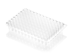
非无菌、透明、聚丙烯 PCR 微孔板
在线联系


 沪公网安备31010102008431号