
 EasySep™小鼠TIL(CD45)正选试剂盒


产品号 #03800
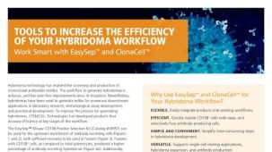
杂交瘤制备全流程解决方案
若您需要咨询产品或有任何技术问题,请通过官方电话 400 885 9050 或邮箱 info.cn@stemcell.com 与我们联系。
杂交瘤制备全流程解决方案
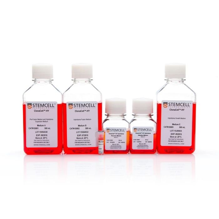
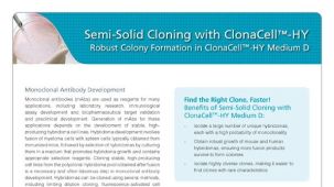
使用ClonaCell™-HY杂交瘤试剂盒中包含的培养基和试剂,完成杂交瘤开发及单克隆抗体生产的所有步骤:
• ClonaCell™-HY培养基A用于骨髓瘤和杂交瘤培养
• ClonaCell™-HY培养基B用于杂交瘤融合
• ClonaCell™-HY培养基C用于杂交瘤融合后恢复
• ClonaCell™-HY培养基D用于杂交瘤筛选与克隆
• ClonaCell™-HY培养基E用于杂交瘤生长
• ClonaCell™-HY PEG用于支持杂交瘤融合
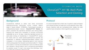
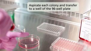
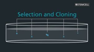
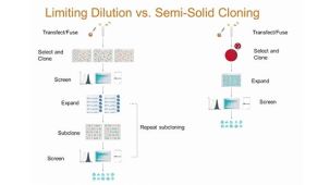
ClonaCell™-HY方法采用基于甲基纤维素的半固体选择性培养基,将杂交瘤筛选和克隆合二为一。在半固体培养基中,单个亲代杂交瘤克隆及其子代在生长形成独立克隆的过程中保持聚集状态。这避免了因生长较快的细胞过度生长而导致稀有克隆丢失的情况,这种情况可能发生在液体培养基筛选过程中。杂交瘤克隆可通过人工或仪器轻松地从半固体培养基中挑取,并分散到液体培养基中进行筛选和扩增。
该试剂盒已验证可用于小鼠和大鼠杂交瘤的构建和单克隆抗体的生产,并兼容多种宿主物种(包括人、小鼠、大鼠和仓鼠)的淋巴细胞,可用于杂交瘤的生产、克隆和扩增。为方便起见,该试剂盒各组分也可单独购买。
分类
半固体培养基,专用培养基
细胞类型
杂交瘤细胞
种属
小鼠,其他物种,大鼠
应用
细胞培养,杂交瘤制备
品牌
ClonaCell
研究领域
抗体制备,细胞系制备,杂交瘤制备
请在《产品说明书》中查找相关支持信息和使用说明,或浏览下方更多实验方案。
本产品专为以下研究领域设计,适用于工作流程中的高亮阶段。探索这些工作流程,了解更多我们为各研究领域提供的其他配套产品。
| 物种 | 其它物种, 大鼠, 小鼠 |
|---|
骨髓瘤与杂交瘤培养基(含血清)
杂交瘤融合培养基(无血清)
杂交瘤融合恢复培养基(含血清)
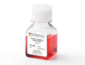
含HAT的杂交瘤筛选和克隆用半固体培养基(含血清))
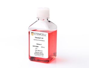
含次黄嘌呤和胸苷的杂交瘤细胞生长培养基(含血清)
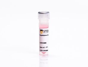
聚乙二醇杂交瘤融合试剂
在线联系


 沪公网安备31010102008431号