Porayette P et al. (DEC 2007)
Biochemical and biophysical research communications 364 3 522--527
Amyloid-?? precursor protein expression and modulation in human embryonic stem cells: A novel role for human chorionic gonadotropin
The amyloid-beta precursor protein (AbetaPP) is a ubiquitously expressed adhesion and neuritogenic protein whose processing has previously been shown to be regulated by reproductive hormones including the gonadotropin luteinizing hormone (LH) in human neuroblastoma cells. We report for the first time the expression of AbetaPP in human embryonic stem (hES) cells at the mRNA and protein levels. Using N- and C-terminal antibodies against AbetaPP,we detected both the mature and immature forms of AbetaPP as well as truncated variants ( approximately 53kDa,47kDa,and 29kDa) by immunoblot analyses. Expression of AbetaPP is regulated by both the stemness of the cells and pregnancy-associated hormones. Addition of human chorionic gonadotropin,the fetal equivalent of LH that is dramatically elevated during pregnancy,markedly increased the expression of all AbetaPP forms. These results indicate a critical molecular signaling link between the hormonal environment of pregnancy and the expression of AbetaPP in hES cells that is suggestive of an important function for this protein during early human embryogenesis prior to the formation of neural precursor cells.
View Publication
产品类型:
产品号#:
05850
05857
05870
05875
85850
85857
85870
85875
产品名:
mTeSR™1
mTeSR™1
Q. Haas et al. ( 2022)
Frontiers in immunology 13 996746
Siglec-7 represents a glyco-immune checkpoint for non-exhausted effector memory CD8+ T cells with high functional and metabolic capacities.
While inhibitory Siglec receptors are known to regulate myeloid cells,less is known about their expression and function in lymphocytes subsets. Here we identified Siglec-7 as a glyco-immune checkpoint expressed on non-exhausted effector memory CD8+ T cells that exhibit high functional and metabolic capacities. Seahorse analysis revealed higher basal respiration and glycolysis levels of Siglec-7+ CD8+ T cells in steady state,and particularly upon activation. Siglec-7 polarization into the T cell immune synapse was dependent on sialoglycan interactions in trans and prevented actin polarization and effective T cell responses. Siglec-7 ligands were found to be expressed on both leukemic stem cells and acute myeloid leukemia (AML) cells suggesting the occurrence of glyco-immune checkpoints for Siglec-7+ CD8+ T cells,which were found in patients' peripheral blood and bone marrow. Our findings project Siglec-7 as a glyco-immune checkpoint and therapeutic target for T cell-driven disorders and cancer.
View Publication
产品类型:
产品号#:
17953
17953RF
100-0710
产品名:
EasySep™人CD8+ T细胞分选试剂盒
RoboSep™ 人CD8+ T细胞分选试剂盒
EasySep™人CD8+ T细胞分选试剂盒
Preuss E et al. ( 2013)
The Journal of biological chemistry 288 49 35287--35296
Pan-mammalian target of rapamycin (mTOR) inhibitor AZD8055 primes rhabdomyosarcoma cells for ABT-737-induced apoptosis by down-regulating Mcl-1 protein.
The PI3K/mammalian Target of Rapamycin (mTOR) pathway is often aberrantly activated in rhabdomyosarcoma (RMS) and represents a promising therapeutic target. Recent evaluation of AZD8055,an ATP-competitive mTOR inhibitor,by the Preclinical Pediatric Testing Program showed in vivo antitumor activity against childhood solid tumors,including RMS. Therefore,in the present study,we searched for AZD8055-based combination therapies. Here,we identify a new synergistic lethality of AZD8055 together with ABT-737,a BH3 mimetic that antagonizes Bcl-2,Bcl-xL,and Bcl-w but not Mcl-1. AZD8055 and ABT-737 cooperate to induce apoptosis in alveolar and embryonal RMS cells in a highly synergistic fashion (combination index textless 0.2). Synergistic induction of apoptosis by AZD8055 and ABT-737 is confirmed on the molecular level,as AZD8055 and ABT-737 cooperate to trigger loss of mitochondrial membrane potential,activation of caspases,and caspase-dependent apoptosis that is blocked by the pan-caspase inhibitor Z-VAD-fmk. Similar to AZD8055,the PI3K/mTOR inhibitor NVP-BEZ235,the PI3K inhibitor NVP-BKM120 and Akt inhibitor synergize with ABT-737 to trigger apoptosis,whereas no cooperativity is found for the mTOR complex 1 inhibitor RAD001. Interestingly,molecular studies reveal a correlation between the ability of different PI3K/mTOR inhibitors to potentiate ABT-737-induced apoptosis and to suppress Mcl-1 protein levels. Importantly,knockdown of Mcl-1 increases ABT-737-induced apoptosis similar to AZD8055/ABT-737 cotreatment. This indicates that AZD8055-mediated suppression of Mcl-1 protein plays an important role in the synergistic drug interaction. By identifying a novel synergistic interaction of AZD8055 and ABT-737,our findings have important implications for the development of molecular targeted therapies for RMS.
View Publication
产品类型:
产品号#:
73002
73004
产品名:
AZD8055
AZD8055
Bhise NS et al. (DEC 2013)
International Journal of Nanomedicine 8 4641--4658
Evaluating the potential of poly(beta-amino ester) nanoparticles for reprogramming human fibroblasts to become induced pluripotent stem cells
BACKGROUND: Gene delivery can potentially be used as a therapeutic for treating genetic diseases,including neurodegenerative diseases,as well as an enabling technology for regenerative medicine. A central challenge in many gene delivery applications is having a safe and effective delivery method. We evaluated the use of a biodegradable poly(beta-amino ester) nanoparticle-based nonviral protocol and compared this with an electroporation-based approach to deliver episomal plasmids encoding reprogramming factors for generation of human induced pluripotent stem cells (hiPSCs) from human fibroblasts.backslashnbackslashnMETHODS: A polymer library was screened to identify the polymers most promising for gene delivery to human fibroblasts. Feeder-independent culturing protocols were developed for nanoparticle-based and electroporation-based reprogramming. The cells reprogrammed by both polymeric nanoparticle-based and electroporation-based nonviral methods were characterized by analysis of pluripotency markers and karyotypic stability. The hiPSC-like cells were further differentiated toward the neural lineage to test their potential for neurodegenerative retinal disease modeling.backslashnbackslashnRESULTS: 1-(3-aminopropyl)-4-methylpiperazine end-terminated poly(1,4-butanediol diacry-late-co-4-amino-1-butanol) polymer (B4S4E7) self-assembled with plasmid DNA to form nanoparticles that were more effective than leading commercially available reagents,including Lipofectamine® 2000,FuGENE® HD,and 25 kDa branched polyethylenimine,for nonviral gene transfer. B4S4E7 nanoparticles showed effective gene delivery to IMR-90 human primary fibroblasts and to dermal fibroblasts derived from a patient with retinitis pigmentosa,and enabled coexpression of exogenously delivered genes,as is needed for reprogramming. The karyotypically normal hiPSC-like cells generated by conventional electroporation,but not by poly(beta-amino ester) reprogramming,could be differentiated toward the neuronal lineage,specifically pseudostratified optic cups.backslashnbackslashnCONCLUSION: This study shows that certain nonviral reprogramming methods may not necessarily be safer than viral approaches and that maximizing exogenous gene expression of reprogramming factors is not sufficient to ensure successful reprogramming.
View Publication
产品类型:
产品号#:
05850
05857
05870
05875
85850
85857
85870
85875
产品名:
mTeSR™1
mTeSR™1
Guia S et al. (MAY 2008)
Blood 111 10 5008--16
A role for interleukin-12/23 in the maturation of human natural killer and CD56+ T cells in vivo.
Natural killer (NK) cells have been originally defined by their naturally occurring" effector function. However�
View Publication
产品类型:
产品号#:
15025
15065
产品名:
RosetteSep™人NK细胞富集抗体混合物
RosetteSep™人NK细胞富集抗体混合物
Kleinstreuer NC et al. (NOV 2011)
Toxicology and Applied Pharmacology 257 1 111--121
Identifying developmental toxicity pathways for a subset of ToxCast chemicals using human embryonic stem cells and metabolomics
Metabolomics analysis was performed on the supernatant of human embryonic stem (hES) cell cultures exposed to a blinded subset of 11 chemicals selected from the chemical library of EPA's ToxCast™ chemical screening and prioritization research project. Metabolites from hES cultures were evaluated for known and novel signatures that may be indicative of developmental toxicity. Significant fold changes in endogenous metabolites were detected for 83 putatively annotated mass features in response to the subset of ToxCast chemicals. The annotations were mapped to specific human metabolic pathways. This revealed strong effects on pathways for nicotinate and nicotinamide metabolism,pantothenate and CoA biosynthesis,glutathione metabolism,and arginine and proline metabolism pathways. Predictivity for adverse outcomes in mammalian prenatal developmental toxicity studies used ToxRefDB and other sources of information,including Stemina Biomarker Discovery's predictive DevTox® model trained on 23 pharmaceutical agents of known developmental toxicity and differing potency. The model initially predicted developmental toxicity from the blinded ToxCast compounds in concordance with animal data with 73% accuracy. Retraining the model with data from the unblinded test compounds at one concentration level increased the predictive accuracy for the remaining concentrations to 83%. These preliminary results on a 11-chemical subset of the ToxCast chemical library indicate that metabolomics analysis of the hES secretome provides information valuable for predictive modeling and mechanistic understanding of mammalian developmental toxicity.
View Publication
产品类型:
产品号#:
05850
05857
05870
05875
85850
85857
85870
85875
产品名:
mTeSR™1
mTeSR™1
Kriz V et al. (NOV 2006)
The Journal of biological chemistry 281 45 34484--91
The SHB adapter protein is required for normal maturation of mesoderm during in vitro differentiation of embryonic stem cells.
Definitive mesoderm arises from a bipotent mesendodermal population,and to study processes controlling its development at this stage,embryonic stem (ES) cells can be employed. SHB (Src homology 2 protein in beta-cells) is an adapter protein previously found to be involved in ES cell differentiation to mesoderm. To further study the role of SHB in this context,we have established ES cell lines deficient for one (SHB+/-) or both SHB alleles (SHB-/-). Differentiating embryoid bodies (EBs) derived from these ES cell lines were used for gene expression analysis. Alternatively,EBs were stained for the blood vessel marker CD31. For hematopoietic differentiation,EBs were differentiated in methylcellulose. SHB-/- EBs exhibited delayed down-regulation of the early mesodermal marker Brachyury. Later mesodermal markers relatively specific for the hematopoietic,vascular,and cardiac lineages were expressed at lower levels on day 6 or 8 of differentiation in EBs lacking SHB. The expression of vascular endothelial growth factor receptor-2 and fibroblast growth factor receptor-1 was also reduced in SHB-/- EBs. SHB-/- EBs demonstrated impaired blood vessel formation after vascular endothelial growth factor stimulation. In addition,the SHB-/- ES cells formed fewer blood cell colonies than SHB+/+ ES cells. It is concluded that SHB is required for appropriate hematopoietic and vascular differentiation and that delayed down-regulation of Brachyury expression may play a role in this context.
View Publication
产品类型:
产品号#:
03434
03444
产品名:
MethoCult™ GF M3434
MethoCult™ GF M3434
Nika K et al. (MAR 2006)
Molecular and cellular biology 26 5 1806--16
Lipid raft targeting of hematopoietic protein tyrosine phosphatase by protein kinase C theta-mediated phosphorylation.
Protein kinase C theta (PKC theta) is unique among PKC isozymes in its translocation to the center of the immune synapse in T cells and its unique downstream signaling. Here we show that the hematopoietic protein tyrosine phosphatase (HePTP) also accumulates in the immune synapse in a PKC theta-dependent manner upon antigen recognition by T cells and is phosphorylated by PKC theta at Ser-225,which is required for lipid raft translocation. Immune synapse translocation was completely absent in antigen-specific T cells from PKC theta-/- mice. In intact T cells,HePTP-S225A enhanced T-cell receptor (TCR)-induced NFAT/AP-1 transactivation,while the acidic substitution mutant was as efficient as wild-type HePTP. We conclude that HePTP is phosphorylated in the immune synapse by PKC theta and thereby targeted to lipid rafts to temper TCR signaling. This represents a novel mechanism for the active immune synapse recruitment and activation of a phosphatase in TCR signaling.
View Publication
Adipose tissue-derived mesenchymal stem cells have in vivo immunosuppressive properties applicable for the control of the graft-versus-host disease.
Previous studies have shown the relevance of bone marrow-derived MSCs (BM-MSCs) in controlling graft-versus-host disease (GVHD) after allogeneic transplantation. Since adipose tissue-derived MSCs (Ad-MSCs) may constitute a good alternative to BM-MSCs,we have expanded MSCs derived from human adipose tissue (hAd-MSCs) and mouse adipose tissue (mAd-MSCs),investigated the immunoregulatory properties of these cells,and evaluated their capacity to control GVHD in mice. The phenotype and immunoregulatory properties of expanded hAd-MSCs were similar to those of human BM-MSCs. Moreover,hAd-MSCs inhibited the proliferation and cytokine secretion of human primary T cells in response to mitogens and allogeneic T cells. Similarly,ex vivo expanded mAd-MSCs had an equivalent immunophenotype and exerted immunoregulatory properties similar to those of hAd-MSCs. Moreover,the infusion of mAd-MSCs in mice transplanted with haploidentical hematopoietic grafts controlled the lethal GVHD that occurred in control recipient mice. These findings constitute the first experimental proof that Ad-MSCs can efficiently control the GVHD associated with allogeneic hematopoietic transplantation,opening new perspectives for the clinical use of Ad-MSCs.
View Publication
产品类型:
产品号#:
05501
05502
05401
05402
05411
产品名:
MesenCult™ MSC 基础培养基(人)
MesenCult™ MSC 刺激补充剂(人)
MesenCult™ 增殖试剂盒(人)
Ovchinnikov DA et al. (SEP 2014)
Stem cell research 13 2 251--261
Transgenic human ES and iPS reporter cell lines for identification and selection of pluripotent stem cells in vitro
Optimization of pluripotent stem cell expansion and differentiation is facilitated by biological tools that permit non-invasive and dynamic monitoring of pluripotency,and the ability to select for an undifferentiated input cell population. Here we report on the generation and characterisation of clonal human embryonic stem (HES3,H9) and human induced pluripotent stem cell lines (UQEW01i-epifibC11) that have been stably modified with an artificial EOS(C3+) promoter driving expression of EGFP and puromycin resistance-conferring proteins. We show that EGFP expression faithfully reports on the pluripotency status of the cells in these lines and that antibiotic selection allows for an efficient elimination of differentiated cells from the cultures. We demonstrate that the extinction of the expression of the pluripotency reporter during differentiation closely correlates with the decrease in expression of conventional pluripotency markers,such as OCT4 (POU5F1),TRA-1-60 and SSEA4 when screening across conditions with various levels of pluripotency-maintaining or differentiation-inducing signals. We further illustrate the utility of these lines for real-time monitoring of pluripotency in embryoid bodies and microfluidic bioreactors.
View Publication
产品类型:
产品号#:
05850
05857
05870
05875
85850
85857
85870
85875
产品名:
mTeSR™1
mTeSR™1
Robinson M et al. (AUG 2016)
Stem Cell Reviews and Reports 12 4 476--483
Functionalizing Ascl1 with Novel Intracellular Protein Delivery Technology for Promoting Neuronal Differentiation of Human Induced Pluripotent Stem Cells
Pluripotent stem cells can become any cell type found in the body. Accordingly,one of the major challenges when working with pluripotent stem cells is producing a highly homogenous population of differentiated cells,which can then be used for downstream applications such as cell therapies or drug screening. The transcription factor Ascl1 plays a key role in neural development and previous work has shown that Ascl1 overexpression using viral vectors can reprogram fibroblasts directly into neurons. Here we report on how a recombinant version of the Ascl1 protein functionalized with intracellular protein delivery technology (Ascl1-IPTD) can be used to rapidly differentiate human induced pluripotent stem cells (hiPSCs) into neurons. We first evaluated a range of Ascl1-IPTD concentrations to determine the most effective amount for generating neurons from hiPSCs cultured in serum free media. Next,we looked at the frequency of Ascl1-IPTD supplementation in the media on differentiation and found that one time supplementation is sufficient enough to trigger the neural differentiation process. Ascl1-IPTD was efficiently taken up by the hiPSCs and enabled rapid differentiation into TUJ1-positive and NeuN-positive populations with neuronal morphology after 8 days. After 12 days of culture,hiPSC-derived neurons produced by Ascl1-IPTD treatment exhibited greater neurite length and higher numbers of branch points compared to neurons derived using a standard neural progenitor differentiation protocol. This work validates Ascl1-IPTD as a powerful tool for engineering neural tissue from pluripotent stem cells.
View Publication

 EasySep™小鼠TIL(CD45)正选试剂盒


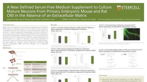 科学海报A New Defined Serum-Free Medium Supplement to Culture Mature Neurons from Primary Embryonic Mouse and Rat CNS
 沪公网安备31010102008431号