Gasparetto M et al. (OCT 2012)
Experimental hematology 40 10 857--66.e5
Varying levels of aldehyde dehydrogenase activity in adult murine marrow hematopoietic stem cells are associated with engraftment and cell cycle status.
Aldehyde dehydrogenase (ALDH) activity is a widely used marker for human hematopoietic stem cells (HSCs),yet its relevance and role in murine HSCs remain unclear. We found that murine marrow cells with a high level of ALDH activity as measured by Aldefluor staining (ALDH(br) cells) do not contain known HSCs or progenitors. In contrast,highly enriched murine HSCs defined by the CD48(-)EPCR(+) and other phenotypes contain two subpopulations,one that stains dimly with Aldefluor (ALDH(dim)) and one that stains at intermediate levels (ALDH(int)). The CD48(-)EPCR(+)ALDH(dim) cells are virtually all in G(0) and yield high levels of engraftment via both intravenous and intrabone routes. In contrast the CD48(-)EPCR(+)ALDH(int) cells are virtually all in G(1),have little intravenous engraftment potential,and yet can engraft long-term after intrabone transplantation. These data demonstrate that Aldefluor staining of unfractionated murine marrow does not identify known HSCs or progenitors. However,varying levels of Aldefluor staining when combined with CD48 and EPCR detection can identify novel populations in murine marrow including a highly enriched population of resting HSCs and a previously unknown HSC population in G(1) with an intravenous engraftment defect.
View Publication
产品类型:
产品号#:
01700
01705
01702
产品名:
ALDEFLUOR™ 试剂盒
ALDEFLUOR™ DEAB试剂, 1.5 mM, 1 mL
ALDEFLUOR™检测缓冲液
Lin H et al. (JAN 2017)
Neuro-oncology 19 1 43--54
Fatty acid oxidation is required for the respiration and proliferation of malignant glioma cells.
BACKGROUND Glioma is the most common form of primary malignant brain tumor in adults,with approximately 4 cases per 100 000 people each year. Gliomas,like many tumors,are thought to primarily metabolize glucose for energy production; however,the reliance upon glycolysis has recently been called into question. In this study,we aimed to identify the metabolic fuel requirements of human glioma cells. METHODS We used database searches and tissue culture resources to evaluate genotype and protein expression,tracked oxygen consumption rates to study metabolic responses to various substrates,performed histochemical techniques and fluorescence-activated cell sorting-based mitotic profiling to study cellular proliferation rates,and employed an animal model of malignant glioma to evaluate a new therapeutic intervention. RESULTS We observed the presence of enzymes required for fatty acid oxidation within human glioma tissues. In addition,we demonstrated that this metabolic pathway is a major contributor to aerobic respiration in primary-cultured cells isolated from human glioma and grown under serum-free conditions. Moreover,inhibiting fatty acid oxidation reduces proliferative activity in these primary-cultured cells and prolongs survival in a syngeneic mouse model of malignant glioma. CONCLUSIONS Fatty acid oxidation enzymes are present and active within glioma tissues. Targeting this metabolic pathway reduces energy production and cellular proliferation in glioma cells. The drug etomoxir may provide therapeutic benefit to patients with malignant glioma. In addition,the expression of fatty acid oxidation enzymes may provide prognostic indicators for clinical practice.
View Publication
产品类型:
产品号#:
05750
05751
产品名:
NeuroCult™ NS-A 基础培养基(人)
NeuroCult™ NS-A 扩增试剂盒(人)
Melkoumian Z et al. (JUN 2010)
Nature biotechnology 28 6 606--10
Synthetic peptide-acrylate surfaces for long-term self-renewal and cardiomyocyte differentiation of human embryonic stem cells.
Human embryonic stem cells (hESCs) have two properties of interest for the development of cell therapies: self-renewal and the potential to differentiate into all major lineages of somatic cells in the human body. Widespread clinical application of hESC-derived cells will require culture methods that are low-cost,robust,scalable and use chemically defined raw materials. Here we describe synthetic peptide-acrylate surfaces (PAS) that support self-renewal of hESCs in chemically defined,xeno-free medium. H1 and H7 hESCs were successfully maintained on PAS for over ten passages. Cell morphology and phenotypic marker expression were similar for cells cultured on PAS or Matrigel. Cells on PAS retained normal karyotype and pluripotency and were able to differentiate to functional cardiomyocytes on PAS. Finally,PAS were scaled up to large culture-vessel formats. Synthetic,xeno-free,scalable surfaces that support the self-renewal and differentiation of hESCs will be useful for both research purposes and development of cell therapies.
View Publication
产品类型:
产品号#:
05860
05880
05850
05857
05870
05875
85850
85857
85870
85875
产品名:
mTeSR™1
mTeSR™1
Zhu F et al. (SEP 2014)
Stem cells and development 23 17 2119--2125
A modified method for implantation of pluripotent stem cells under the rodent kidney capsule.
Teratoma formation,the standard in vivo pluripotency assay,is also frequently used as a tumorigenicity assay. A common concern in therapeutic stem cell applications is the tumorigenicity potential of a small number of cell impurities in the final product. Estimation of this small number is hampered by the inaccurate methodology of the tumorigenicity assay. Hence,a protocol for tumorigenicity assay that can deliver a defined number of cells,without error introduced by leakage or migration of cells is needed. In this study,we tested our modified transplantation method that allows for transplant of small numbers of pluripotent stem cells (PSCs) under the kidney capsule with minimal cell leakage. A glass capillary with a finely shaped tip and an attached mouth pipette was used to inject PSCs into the rodent kidney capsule. H9 embryonic and induced PSCs were tagged with Fluc and green fluorescence protein reporter genes and divided in different cell doses for transplantation. Bioluminescence imaging (BLI) on the day of surgery showed that the cell signal was confined to the kidney and signal intensity correlated with increasing transplant cell numbers. The overall cell leakage rate was 17% and the rodent survival rate was 96%. Teratoma formation was observed in rodents transplanted with cell numbers between 1 × 10(5)-2 × 10(6). We conclude that this modified procedure for transplanting PSCs under the kidney capsule allows for transplantation of a defined number of PSCs with significant reduction of error associated with cell leakage from the transplant site.
View Publication
产品类型:
产品号#:
05850
05857
05870
05875
85850
85857
85870
85875
产品名:
mTeSR™1
mTeSR™1
Zhu X et al. (SEP 2014)
Sci Rep 4 6420
An efficient genotyping method for genome-modified animals and human cells generated with CRISPR/Cas9 system
The rapid generation of various species and strains of laboratory animals using CRISPR/Cas9 technology has dramatically accelerated the interrogation of gene function in vivo. So far,the dominant approach for genotyping of genome-modified animals has been the T7E1 endonuclease cleavage assay. Here,we present a polyacrylamide gel electrophoresis-based (PAGE) method to genotype mice harboring different types of indel mutations. We developed 6 strains of genome-modified mice using CRISPR/Cas9 system,and utilized this approach to genotype mice from F0 to F2 generation,which included single and multiplexed genome-modified mice. We also determined the maximal detection sensitivity for detecting mosaic DNA using PAGE-based assay as 0.5%. We further applied PAGE-based genotyping approach to detect CRISPR/Cas9-mediated on- and off-target effect in human 293T and induced pluripotent stem cells (iPSCs). Thus,PAGE-based genotyping approach meets the rapidly increasing demand for genotyping of the fast-growing number of genome-modified animals and human cell lines created using CRISPR/Cas9 system or other nuclease systems such as TALEN or ZFN.
View Publication
产品类型:
产品号#:
05850
05857
05870
05875
85850
85857
85870
85875
产品名:
mTeSR™1
mTeSR™1
Sancho-Martinez I et al. (FEB 2016)
Nature communications 7 10743
Establishment of human iPSC-based models for the study and targeting of glioma initiating cells.
Glioma tumour-initiating cells (GTICs) can originate upon the transformation of neural progenitor cells (NPCs). Studies on GTICs have focused on primary tumours from which GTICs could be isolated and the use of human embryonic material. Recently,the somatic genomic landscape of human gliomas has been reported. RTK (receptor tyrosine kinase) and p53 signalling were found dysregulated in ∼90% and 86% of all primary tumours analysed,respectively. Here we report on the use of human-induced pluripotent stem cells (hiPSCs) for modelling gliomagenesis. Dysregulation of RTK and p53 signalling in hiPSC-derived NPCs (iNPCs) recapitulates GTIC properties in vitro. In vivo transplantation of transformed iNPCs leads to highly aggressive tumours containing undifferentiated stem cells and their differentiated derivatives. Metabolic modulation compromises GTIC viability. Last,screening of 101 anti-cancer compounds identifies three molecules specifically targeting transformed iNPCs and primary GTICs. Together,our results highlight the potential of hiPSCs for studying human tumourigenesis.
View Publication
产品类型:
产品号#:
05850
05857
05870
05875
85850
85857
85870
85875
产品名:
mTeSR™1
mTeSR™1
(Oct 2024)
Nature Communications 15
Reassessment of marker genes in human induced pluripotent stem cells for enhanced quality control
Human induced pluripotent stem cells (iPSCs) have great potential in research,but pluripotency testing faces challenges due to non-standardized methods and ambiguous markers. Here,we use long-read nanopore transcriptome sequencing to discover 172 genes linked to cell states not covered by current guidelines. We validate 12 genes by qPCR as unique markers for specific cell fates: pluripotency (CNMD,NANOG,SPP1),endoderm (CER1,EOMES,GATA6),mesoderm (APLNR,HAND1,HOXB7),and ectoderm (HES5,PAMR1,PAX6). Using these genes,we develop a machine learning-based scoring system,“hiPSCore”,trained on 15 iPSC lines and validated on 10 more. hiPSCore accurately classifies pluripotent and differentiated cells and predicts their potential to become specialized 2D cells and 3D organoids. Our re-evaluation of cell fate marker genes identifies key targets for future studies on cell fate assessment. hiPSCore improves iPSC testing by reducing time,subjectivity,and resource use,thus enhancing iPSC quality for scientific and medical applications. Quality control,including pluripotency testing of human iPSCs lacks standardization. Here,authors identify and validate gene markers to develop the machine learning-based hiPSCore to streamline pluripotency testing and elevate iPSC quality.
View Publication
产品类型:
产品号#:
05230
05310
08581
08582
100-0038
100-0041
100-0195
100-0651
100-0276
100-1130
产品名:
STEMdiff™ 三胚层分化试剂盒
STEMdiff™ 造血试剂盒
STEMdiff™SMADi神经诱导试剂盒
STEMdiff™SMADi神经诱导试剂盒,2套
STEMdiff™中脑神经元分化试剂盒
STEMdiff™ 中脑神经元成熟试剂盒
STEMdiff™带分支结构的肺类器官试剂盒
STEMdiff™ 血管类器官试剂盒
mTeSR™ Plus
mTeSR™ Plus
(Nov 2024)
Frontiers in Immunology 15 6335
Human CD34+-derived plasmacytoid dendritic cells as surrogates for primary pDCs and potential cancer immunotherapy
IntroductionPlasmacytoid dendritic cells (pDCs) are capable of triggering broad immune responses,yet,their scarcity in blood coupled to their reduced functionality in cancer,makes their therapeutic use for in situ activation or vaccination challenging. MethodsWe designed an in vitro differentiation protocol tailored for human pDCs from cord blood (CB) hematopoietic stem cells (HSCs) with StemRegenin 1 (SR-1) and GM-CSF supplementation. Next,we evaluated the identity and function of CB-pDCs compared to human primary pDCs. Furthermore,we tested the potential of CB-pDCs to support anti-tumor immune responses in co-culture with tumor explants from CRC patients. ResultsHere,we report an in vitro differentiation protocol enabling the generation of 200 pDCs per HSC and highlight the role of GM-CSF and SR-1 in CB-pDC differentiation and function. CB-pDCs exhibited a robust resemblance to primary pDCs phenotypically and functionally. Transcriptomic analysis confirmed strong homology at both,baseline and upon TLR9 or TLR7 stimulation. Further,we could confirm the potential of CB-pDCs to promote inflammation in the tumor microenvironment by eliciting cytokines associated with NK and T cell recruitment and function upon TLR7 stimulation ex vivo in patient tumor explants. DiscussionThis study highlights CB-pDCs as surrogates for primary pDCs to investigate their biology and for their potential use as cell therapy in cancer.
View Publication
产品类型:
产品号#:
19055
19055RF
产品名:
EasySep™人NK细胞富集试剂盒
RoboSep™ 人NK细胞富集试剂盒含滤芯吸头
Q. Yin et al. (Nov 2025)
Nature Communications 16
Transcription factor ZNF263 primes human embryonic stem cells for pluripotency dissolution and lineage commitment
Conventional human embryonic stem cells (hESCs) are capable of self-renewal and simultaneously poised for differentiation. But the mechanisms underlying this primed pluripotent state,which endows them with elevated responsiveness to differentiation cues,remain largely underexplored. Especially,little is known about the pivotal transcription factors (TFs) that orchestrate hESCs towards primed pluripotency. Here,we report a function of TF ZNF263 in pluripotency priming. Genetic and functional assays reveal that ZNF263 directly initiates the incipient expression of early differentiation genes and concurrently dampens the core pluripotency circuitry in hESCs,greatly tilting the balance from pluripotency maintenance to lineage priming. Importantly,ZNF263 deficiency markedly impairs pluripotency dissolution and multi-lineage differentiation in hESCs,particularly toward ectoderm. Moreover,single-cell transcriptomic profiling reveals that ZNF263 promotes the priming of cell fate commitment in hESCs,suggesting its indispensable requirement for pluripotency priming and lineage commitment continuum. Together,we demonstrate the role of ZNF263 in establishing the primed pluripotent state in hESCs and facilitating their differentiation into primary germ layer lineages. Human embryonic stem cells are simultaneously capable of self-renewal and poised for differentiation. Here,the authors show a role for the ZNF263 transcription factor promotes primed pluripotency and facilitates differentiation into primary germ layer lineages.
View Publication
ErbB4 Activated p38$$ MAPK Isoform Mediates Early Cardiogenesis Through NKx2.5 in Human Pluripotent Stem Cells
Activation of ErbB4 receptor signaling is instrumental in heart development,lack of which results in embryonic lethality. However,mechanism governing its intracellular signaling remains elusive. Using human pluripotent stem cells,we show that ErbB4 is critical for cardiogenesis whereby its genetic knockdown results in loss of cardiomyocytes. Phospho-proteome profiling and Western blot studies attribute this loss to inactivation of p38$\$ isoform which physically interacts with NKx2.5 and GATA4 transcription factors. Post-cardiomyocyte formation p38$\$/NKx2.5 downregulation is followed by p38$\$/MEF2c upregulation suggesting stage-specific developmental roles of p38 MAPK isoforms. Knockdown of p38$\$ similarly disrupts cardiomyocyte formation in spite of the presence of NKx2.5. Cell fractionation and NKx2.5 phosphorylation studies suggest inhibition of ErbB4-p38$\$ hinders NKx2.5 nuclear translocation during early cardiogenesis. This study reveals a novel pathway that directly links ErbB4 and p38$\$ the transcriptional machinery of NKx2.5-GATA4 complex which is critical for cardiomyocyte formation during mammalian heart development.
View Publication

 EasySep™小鼠TIL(CD45)正选试剂盒


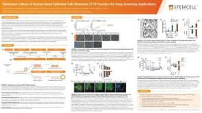 科学海报Optimized Culture of Human Nasal Epithelial Cells Maintains CFTR Function for Drug Screening Applications


 沪公网安备31010102008431号