Bhise NS et al. (DEC 2013)
International Journal of Nanomedicine 8 4641--4658
Evaluating the potential of poly(beta-amino ester) nanoparticles for reprogramming human fibroblasts to become induced pluripotent stem cells
BACKGROUND: Gene delivery can potentially be used as a therapeutic for treating genetic diseases,including neurodegenerative diseases,as well as an enabling technology for regenerative medicine. A central challenge in many gene delivery applications is having a safe and effective delivery method. We evaluated the use of a biodegradable poly(beta-amino ester) nanoparticle-based nonviral protocol and compared this with an electroporation-based approach to deliver episomal plasmids encoding reprogramming factors for generation of human induced pluripotent stem cells (hiPSCs) from human fibroblasts.backslashnbackslashnMETHODS: A polymer library was screened to identify the polymers most promising for gene delivery to human fibroblasts. Feeder-independent culturing protocols were developed for nanoparticle-based and electroporation-based reprogramming. The cells reprogrammed by both polymeric nanoparticle-based and electroporation-based nonviral methods were characterized by analysis of pluripotency markers and karyotypic stability. The hiPSC-like cells were further differentiated toward the neural lineage to test their potential for neurodegenerative retinal disease modeling.backslashnbackslashnRESULTS: 1-(3-aminopropyl)-4-methylpiperazine end-terminated poly(1,4-butanediol diacry-late-co-4-amino-1-butanol) polymer (B4S4E7) self-assembled with plasmid DNA to form nanoparticles that were more effective than leading commercially available reagents,including Lipofectamine® 2000,FuGENE® HD,and 25 kDa branched polyethylenimine,for nonviral gene transfer. B4S4E7 nanoparticles showed effective gene delivery to IMR-90 human primary fibroblasts and to dermal fibroblasts derived from a patient with retinitis pigmentosa,and enabled coexpression of exogenously delivered genes,as is needed for reprogramming. The karyotypically normal hiPSC-like cells generated by conventional electroporation,but not by poly(beta-amino ester) reprogramming,could be differentiated toward the neuronal lineage,specifically pseudostratified optic cups.backslashnbackslashnCONCLUSION: This study shows that certain nonviral reprogramming methods may not necessarily be safer than viral approaches and that maximizing exogenous gene expression of reprogramming factors is not sufficient to ensure successful reprogramming.
View Publication
产品类型:
产品号#:
05850
05857
05870
05875
85850
85857
85870
85875
产品名:
mTeSR™1
mTeSR™1
Guia S et al. (MAY 2008)
Blood 111 10 5008--16
A role for interleukin-12/23 in the maturation of human natural killer and CD56+ T cells in vivo.
Natural killer (NK) cells have been originally defined by their naturally occurring" effector function. However�
View Publication
产品类型:
产品号#:
15025
15065
产品名:
RosetteSep™人NK细胞富集抗体混合物
RosetteSep™人NK细胞富集抗体混合物
Kleinstreuer NC et al. (NOV 2011)
Toxicology and Applied Pharmacology 257 1 111--121
Identifying developmental toxicity pathways for a subset of ToxCast chemicals using human embryonic stem cells and metabolomics
Metabolomics analysis was performed on the supernatant of human embryonic stem (hES) cell cultures exposed to a blinded subset of 11 chemicals selected from the chemical library of EPA's ToxCast™ chemical screening and prioritization research project. Metabolites from hES cultures were evaluated for known and novel signatures that may be indicative of developmental toxicity. Significant fold changes in endogenous metabolites were detected for 83 putatively annotated mass features in response to the subset of ToxCast chemicals. The annotations were mapped to specific human metabolic pathways. This revealed strong effects on pathways for nicotinate and nicotinamide metabolism,pantothenate and CoA biosynthesis,glutathione metabolism,and arginine and proline metabolism pathways. Predictivity for adverse outcomes in mammalian prenatal developmental toxicity studies used ToxRefDB and other sources of information,including Stemina Biomarker Discovery's predictive DevTox® model trained on 23 pharmaceutical agents of known developmental toxicity and differing potency. The model initially predicted developmental toxicity from the blinded ToxCast compounds in concordance with animal data with 73% accuracy. Retraining the model with data from the unblinded test compounds at one concentration level increased the predictive accuracy for the remaining concentrations to 83%. These preliminary results on a 11-chemical subset of the ToxCast chemical library indicate that metabolomics analysis of the hES secretome provides information valuable for predictive modeling and mechanistic understanding of mammalian developmental toxicity.
View Publication
Ovchinnikov DA et al. (SEP 2014)
Stem cell research 13 2 251--261
Transgenic human ES and iPS reporter cell lines for identification and selection of pluripotent stem cells in vitro
Optimization of pluripotent stem cell expansion and differentiation is facilitated by biological tools that permit non-invasive and dynamic monitoring of pluripotency,and the ability to select for an undifferentiated input cell population. Here we report on the generation and characterisation of clonal human embryonic stem (HES3,H9) and human induced pluripotent stem cell lines (UQEW01i-epifibC11) that have been stably modified with an artificial EOS(C3+) promoter driving expression of EGFP and puromycin resistance-conferring proteins. We show that EGFP expression faithfully reports on the pluripotency status of the cells in these lines and that antibiotic selection allows for an efficient elimination of differentiated cells from the cultures. We demonstrate that the extinction of the expression of the pluripotency reporter during differentiation closely correlates with the decrease in expression of conventional pluripotency markers,such as OCT4 (POU5F1),TRA-1-60 and SSEA4 when screening across conditions with various levels of pluripotency-maintaining or differentiation-inducing signals. We further illustrate the utility of these lines for real-time monitoring of pluripotency in embryoid bodies and microfluidic bioreactors.
View Publication
产品类型:
产品号#:
05850
05857
05870
05875
85850
85857
85870
85875
产品名:
mTeSR™1
mTeSR™1
Robinson M et al. (AUG 2016)
Stem Cell Reviews and Reports 12 4 476--483
Functionalizing Ascl1 with Novel Intracellular Protein Delivery Technology for Promoting Neuronal Differentiation of Human Induced Pluripotent Stem Cells
Pluripotent stem cells can become any cell type found in the body. Accordingly,one of the major challenges when working with pluripotent stem cells is producing a highly homogenous population of differentiated cells,which can then be used for downstream applications such as cell therapies or drug screening. The transcription factor Ascl1 plays a key role in neural development and previous work has shown that Ascl1 overexpression using viral vectors can reprogram fibroblasts directly into neurons. Here we report on how a recombinant version of the Ascl1 protein functionalized with intracellular protein delivery technology (Ascl1-IPTD) can be used to rapidly differentiate human induced pluripotent stem cells (hiPSCs) into neurons. We first evaluated a range of Ascl1-IPTD concentrations to determine the most effective amount for generating neurons from hiPSCs cultured in serum free media. Next,we looked at the frequency of Ascl1-IPTD supplementation in the media on differentiation and found that one time supplementation is sufficient enough to trigger the neural differentiation process. Ascl1-IPTD was efficiently taken up by the hiPSCs and enabled rapid differentiation into TUJ1-positive and NeuN-positive populations with neuronal morphology after 8 days. After 12 days of culture,hiPSC-derived neurons produced by Ascl1-IPTD treatment exhibited greater neurite length and higher numbers of branch points compared to neurons derived using a standard neural progenitor differentiation protocol. This work validates Ascl1-IPTD as a powerful tool for engineering neural tissue from pluripotent stem cells.
View Publication
产品类型:
产品号#:
05832
05833
05838
05872
05873
05940
07174
07180
07183
07190
27147
07191
36254
07930
07931
07940
07955
07956
07959
07954
27845
27945
27840
27865
27940
27965
05835
05839
08581
08582
100-0483
100-0484
100-1061
07952
100-0763
100-0485
100-1077
产品名:
STEMdiff™ 神经花环选择试剂
STEMdiff™神经前体细胞培养基
STEMdiff™神经祖细胞冻存液
Vitronectin XF™
CellAdhere™ 稀释缓冲液
DMEM/F-12 with 15 mM HEPES
CryoStor® CS10
CryoStor® CS10
CryoStor® CS10
CryoStor® CS10
CryoStor® CS10
STEMdiff™ 神经诱导培养基
STEMdiff™ 神经诱导培养基
STEMdiff™SMADi神经诱导试剂盒
STEMdiff™SMADi神经诱导试剂盒,2套
Hausser Scientificᵀᴹ 明线血球计数板
ReLeSR™
CryoStor® CS10
CryoStor® CS10
Vitronectin XF™
温和细胞解离试剂
ReLeSR™
Kim J et al. (NOV 2013)
Stem Cell Research 11 3 978--989
Alginate microcapsule as a 3D platform for the efficient differentiation of human embryonic stem cells to dopamine neurons
Human embryonic stem cells (hESCs) are emerging as an attractive alternative source for cell replacement therapy since the cells can be expanded in culture indefinitely and differentiated into any cell types in the body. In order to optimize cell-to-cell interaction,cell proliferation and differentiation into specific lineages as well as tissue organization,it is important to provide a microenvironment for the hESCs which mimics the stem cell niche. One approach is to provide a three-dimensional (3D) environment such as encapsulation. We present an approach to culture and differentiate hESCs into midbrain dopamine (mdDA) neurons in a 3D microenvironment using alginate microcapsules for the first time. A detailed gene and protein expression analysis during neuronal differentiation showed an increased gene and protein expression of various specific DA neuronal markers,particularly tyrosine hydroxylase (TH) by textgreater100 folds after 2weeks and at least 50% higher expression after 4weeks respectively,compared to cells differentiated under conventional two-dimensional (2D) platform. The encapsulated TH+ cells co-expressed mdDA neuronal markers,forkhead box protein A-2 (FOXA2) and pituitary homeobox-3 (PITX3) after 4weeks and secreted approximately 60pg/ml/106 cells higher DA level when induced. We propose that the 3D platform facilitated an early onset of DA neuronal generation compared to that with conventional 2D system which also secretes more DA under potassium-induction. It is a very useful model to study the proliferation and directed differentiation of hESCs to various lineages,particularly to mdDA neurons. This 3D system also allows the separation of feeder cells from hESCs during the process of differentiation and also has potential for immune-isolation during transplantation studies. ?? 2013 Elsevier B.V.
View Publication
产品类型:
产品号#:
05850
05857
05870
05875
07923
85850
85857
85870
85875
产品名:
Dispase (1 U/mL)
mTeSR™1
mTeSR™1
Nagano M et al. (JUL 2007)
Blood 110 1 151--60
Identification of functional endothelial progenitor cells suitable for the treatment of ischemic tissue using human umbilical cord blood.
Umbilical cord blood (UCB) has been used as a potential source of various kinds of stem cells,including hematopoietic stem cells,mesenchymal stem cells,and endothelial progenitor cells (EPCs),for a variety of cell therapies. Recently,EPCs were introduced for restoring vascularization in ischemic tissues. An appropriate procedure for isolating EPCs from UCB is a key issue for improving therapeutic efficacy and eliminating the unexpected expansion of nonessential cells. Here we report a novel method for isolating EPCs from UCB by a combination of negative immunoselection and cell culture techniques. In addition,we divided EPCs into 2 subpopulations according to the aldehyde dehydrogenase (ALDH) activity. We found that EPCs with low ALDH activity (Alde-Low) possess a greater ability to proliferate and migrate compared to those with high ALDH activity (Alde-High). Moreover,hypoxia-inducible factor proteins are up-regulated and VEGF,CXCR4,and GLUT-1 mRNAs are increased in Alde-Low EPCs under hypoxic conditions,while the response was not significant in Alde-High EPCs. In fact,the introduction of Alde-Low EPCs significantly reduced tissue damage in ischemia in a mouse flap model. Thus,the introduction of Alde-Low EPCs may be a potential strategy for inducing rapid neovascularization and subsequent regeneration of ischemic tissues.
View Publication
产品类型:
产品号#:
01700
01705
15128
15168
01702
产品名:
ALDEFLUOR™ 试剂盒
ALDEFLUOR™ DEAB试剂, 1.5 mM, 1 mL
RosetteSep™人间充质干细胞富集抗体混合物
RosetteSep™人间充质干细胞富集抗体混合物
ALDEFLUOR™检测缓冲液
Jones C et al. (MAY 2004)
Cancer research 64 9 3037--45
Expression profiling of purified normal human luminal and myoepithelial breast cells: identification of novel prognostic markers for breast cancer.
The normal duct-lobular system of the breast is lined by two epithelial cell types,inner luminal secretory cells and outer contractile myoepithelial cells. We have generated comprehensive expression profiles of the two normal cell types,using immunomagnetic cell separation and gene expression microarray analysis. The cell-type specificity was confirmed at the protein level by immunohistochemistry in normal breast tissue. New prognostic markers for survival were identified when the luminal- and myoepithelial-specific molecules were evaluated on breast tumor tissue microarrays. Nuclear expression of luminal epithelial marker galectin 3 correlated with a shorter overall survival in these patients,and the expression of SPARC (osteonectin),a myoepithelial marker,was an independent marker of poor prognosis in breast cancers as a whole. These data provide a framework for the interpretation of breast cancer molecular profiling experiments,the identification of potential new diagnostic markers,and development of novel indicators of prognosis.
View Publication
产品类型:
产品号#:
01434
产品名:
H. Cai et al. (Jan 2026)
International Journal of Molecular Sciences 27 1
NGR1 Pretreatment Enhances the Therapeutic Efficacy of Transplanting Cardiomyocytes Derived from Human Induced Pluripotent Stem Cells for Myocardial Infarction
Human induced pluripotent stem cells (hiPSCs) offer significant potential for differentiation and research applications in cardiovascular diseases. When induced differentiated hiPSC-derived cardiomyocytes (hiPSC-CMs) are transplanted into the infarcted myocardial region,they exhibit extremely low survival rates and unsatisfactory therapeutic effects due to ischemia,hypoxia,and immune inflammation in the surrounding environment. To address this issue,we used Panax notoginseng saponin R1 (NGR1),which has demonstrated significant protective effects in prior research,to pretreat hiPSC-CMs before transplantation. Utilizing an in vitro H2O2 oxidative stress model and a nude mouse myocardial infarction (MI) model,we investigated the mechanism through which NGR1 pretreatment enhances the therapeutic efficacy of hiPSC-CM transplantation. The results revealed that the hiPSC-CMs expressed cTnT. NGR1 did not promote the proliferation of hiPSC-CMs but instead induced elevated levels of p-Akt protein in these cells. Compared to hiPSC-CM transplantation alone,transplantation of hiPSC-CMs pretreated with NGR1 exhibited higher ejection fraction (EF) and fractional shortening (FS) values,along with reduced infarct size and collagen deposition. Additionally,there were more HNA-positive cardiomyocytes in the cardiac tissue,fewer TUNEL-positive signals,and increased VWF-positive and Lyve1-positive signals. Furthermore,the gene expression levels of VEGFC,IGF-1,and SDF-1 were higher. Therefore,NGR1 pretreatment improves the survival of transplanted hiPSC-CMs in tissues,reduces myocardial apoptosis,enhances cardiac function,decreases infarct size and collagen deposition,promotes angiogenesis and lymphangiogenesis,and stimulates paracrine secretion.
View Publication
产品类型:
产品号#:
85850
85857
产品名:
mTeSR™1
mTeSR™1
(Nov 2024)
International Journal of Molecular Sciences 25 22
The Generation of Genetically Engineered Human Induced Pluripotent Stem Cells Overexpressing IFN-? for Future Experimental and Clinically Oriented Studies
Induced pluripotent stem cells (iPSCs) can be generated from various adult cells,genetically modified and differentiated into diverse cell populations. Type I interferons (IFN-Is) have multiple immunotherapeutic applications; however,their systemic administration can lead to severe adverse outcomes. One way of overcoming the limitation is to introduce cells able to enter the site of pathology and to produce IFN-Is locally. As a first step towards the generation of such cells,here,we aimed to generate human iPSCs overexpressing interferon-beta (IFNB,IFNB-iPSCs). IFNB-iPSCs were obtained by CRISPR/Cas9 editing of the previously generated iPSC line K7-4Lf. IFNB-iPSCs overexpressed IFNB RNA and produced a functionally active IFN-?. The cells displayed typical iPSC morphology and expressed pluripotency markers. Following spontaneous differentiation,IFNB-iPSCs formed embryoid bodies and upregulated endoderm,mesoderm,and some ectoderm markers. However,an upregulation of key neuroectoderm markers,PAX6 and LHX2,was compromised. A negative effect of IFN-? on iPSC neuroectoderm differentiation was confirmed in parental iPSCs differentiated in the presence of a recombinant IFN-?. The study describes new IFN-?-producing iPSC lines suitable for the generation of various types of IFN-?-producing cells for future experimental and clinical applications,and it unravels an inhibitory effect of IFN-? on stem cell neuroectoderm differentiation.
View Publication
产品类型:
产品号#:
85850
85857
产品名:
mTeSR™1
mTeSR™1
Zhu Y et al. (JAN 2013)
PLoS ONE 8 1 e54552
Three-Dimensional Neuroepithelial Culture from Human Embryonic Stem Cells and Its Use for Quantitative Conversion to Retinal Pigment Epithelium
A goal in human embryonic stem cell (hESC) research is the faithful differentiation to given cell types such as neural lineages. During embryonic development,a basement membrane surrounds the neural plate that forms a tight,apico-basolaterally polarized epithelium before closing to form a neural tube with a single lumen. Here we show that the three-dimensional epithelial cyst culture of hESCs in Matrigel combined with neural induction results in a quantitative conversion into neuroepithelial cysts containing a single lumen. Cells attain a defined neuroepithelial identity by 5 days. The neuroepithelial cysts naturally generate retinal epithelium,in part due to IGF-1/insulin signaling. We demonstrate the utility of this epithelial culture approach by achieving a quantitative production of retinal pigment epithelial (RPE) cells from hESCs within 30 days. Direct transplantation of this RPE into a rat model of retinal degeneration without any selection or expansion of the cells results in the formation of a donor-derived RPE monolayer that rescues photoreceptor cells. The cyst method for neuroepithelial differentiation of pluripotent stem cells is not only of importance for RPE generation but will also be relevant to the production of other neuronal cell types and for reconstituting complex patterning events from three-dimensional neuroepithelia.
View Publication

 EasySep™小鼠TIL(CD45)正选试剂盒


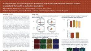 科学海报A Fully Defined Animal Component Free Medium for Efficient Differentiation of Human Pluripotent Stem Cells to Definitive Endoderm
 沪公网安备31010102008431号