Kumar S et al. ( 2016)
Stem Cells International 2016 1--20
Utility of Lymphoblastoid Cell Lines for Induced Pluripotent Stem Cell Generation
A large number of EBV immortalized LCLs have been generated and maintained in genetic/epidemiological studies as a perpetual source of DNA and as a surrogate in vitro cell model. Recent successes in reprograming LCLs into iPSCs have paved the way for generating more relevant in vitro disease models using this existing bioresource. However,the overall reprogramming efficiency and success rate remain poor and very little is known about the mechanistic changes that take place at the transcriptome and cellular functional level during LCL-to-iPSC reprogramming. Here,we report a new optimized LCL-to-iPSC reprogramming protocol using episomal plasmids encoding pluripotency transcription factors and mouse p53DD (p53 carboxy-terminal dominant-negative fragment) and commercially available reprogramming media. We achieved a consistently high reprogramming efficiency and 100% success rate using this optimized protocol. Further,we investigated the transcriptional changes in mRNA and miRNA levels,using FC-abs ≥ 2.0 and FDR ≤ 0.05 cutoffs; 5,228 mRNAs and 77 miRNAs were differentially expressed during LCL-to-iPSC reprogramming. The functional enrichment analysis of the upregulated genes and activation of human pluripotency pathways in the reprogrammed iPSCs showed that the generated iPSCs possess transcriptional and functional profiles very similar to those of human ESCs.
View Publication
产品类型:
产品号#:
05850
05857
05870
05875
05910
85850
85857
85870
85875
产品名:
mTeSR™1
mTeSR™1
M. Boyer et al. (jan 2020)
Scientific reports 10 1 1612
Circulating Tumor Cell Detection and Polyomavirus Status in Merkel Cell Carcinoma.
The incidence of Merkel cell carcinoma (MCC),a rare and highly metastatic skin malignancy,has sharply increased in the last decade. Clinical biomarkers are urgently needed for MCC prognosis,treatment response monitoring,and early diagnosis of relapse. The clinical interest of circulating tumors cells (CTCs) has been validated in many solid cancers. The aim of this study was to compare CTC detection and characterization in blood samples of patients with MCC using the CellSearch System and the RosetteSep -DEPArray workflow,an innovative procedure to enrich,detect and isolate single CTCs. In preliminary experiments (using spiked MCC cell lines) both methods allowed detecting very few MCC cells. In blood samples from 19 patients with MCC at different stages,CellSearch detected MCC CTCs in 26{\%} of patients,and the R-D workflow in 42{\%} of patients. The detection of CTC-positive patients increased to 52{\%} by the cumulative positivity rate of both methodologies. Moreover,Merkel cell polyomavirus DNA,involved in MCC oncogenesis,was detected in tumor biopsies,but not in all single CTCs from the same patient,reflecting the tumor heterogeneity. Our data demonstrate the possibility to detect,isolate and characterize CTCs in patients with MCC using two complementary approaches.
View Publication
BACKGROUND: Multiple measures of endothelial progenitor cells (EPCs) have been described,but there has been limited study of the comparability of these assays. We sought to determine the reproducibility of and correlation between alternative EPC assay methodologies. METHODS: We simultaneously assessed EPC numbers in 140 patients undergoing cardiac catheterization using the 2 most commonly used culture techniques: endothelial cell outgrowth and colony-forming unit (CFU). In the final 77 patients,EPCs were also identified on the basis of cell surface marker expression (CD133,CD34,and vascular endothelial growth factor receptor-2 [VEGFR-2]) and aldehyde dehydrogenase (ALDH) activity. RESULTS: Endothelial progenitor cell enumeration based on fluorescence activated cell sorting was more precise than culture assays. There was limited correlation between EPC numbers determined using the 2 common culture-based assays; however,endothelial CFUs correlated with VEGFR-2 and CD34/VEGFR-2-expressing cells. Endothelial progenitor cells defined by expression of CD133,CD34,CD133/CD34,and ALDH activity correlated with each other,but not with VEGFR-2(+) cells. CONCLUSIONS: Endothelial progenitor cells can be broadly classified into 2 classes: VEGFR-2-expressing cells,which give rise to endothelial CFUs,and CD133/CD34 or ALDH(br) cells. These observations underscore the need for better assay standardization and a more precise definition of EPCs in cell therapy research.
View Publication
产品类型:
产品号#:
01700
01705
01701
01702
产品名:
ALDEFLUOR™ 试剂盒
ALDEFLUOR™ DEAB试剂, 1.5 mM, 1 mL
ALDEFLUOR™检测缓冲液
Wu K et al. (JAN 2011)
The Journal of biological chemistry 286 3 2132--42
Cell fate determination factor Dachshund reprograms breast cancer stem cell function.
The cell fate determination factor Dachshund was cloned as a dominant inhibitor of the hyperactive epidermal growth factor receptor ellipse. The expression of Dachshund is lost in human breast cancer associated with poor prognosis. Breast tumor-initiating cells (TIC) may contribute to tumor progression and therapy resistance. Here,endogenous DACH1 was reduced in breast cancer cell lines with high expression of TIC markers and in patient samples of the basal breast cancer phenotype. Re-expression of DACH1 reduced new tumor formation in serial transplantations in vivo,reduced mammosphere formation,and reduced the proportion of CD44(high)/CD24(low) breast tumor cells. Conversely,lentiviral shRNA to DACH1 increased the breast (B)TIC population. Genome-wide expression studies of mammary tumors demonstrated DACH1 repressed a molecular signature associated with stem cells (SOX2,Nanog,and KLF4) and genome-wide ChIP-seq analysis identified DACH1 binding to the promoter of the Nanog,KLF4,and Lin28 genes. KLF4/c-Myc and Oct4/Sox2 antagonized DACH1 repression of BTIC. Mechanistic studies demonstrated DACH1 directly repressed the Nanog and Sox2 promoters via a conserved domain. Endogenous DACH1 regulates BTIC in vitro and in vivo.
View Publication
产品类型:
产品号#:
01700
01705
01702
产品名:
ALDEFLUOR™ 试剂盒
ALDEFLUOR™ DEAB试剂, 1.5 mM, 1 mL
ALDEFLUOR™检测缓冲液
Wang TH et al. ( 2000)
Cancer 88 11 2619--2628
Paclitaxel-induced cell death: where the cell cycle and apoptosis come together.
BACKGROUND: Compelling evidence indicates that paclitaxel kills cancer cells through the induction of apoptosis. Paclitaxel binds microtubules and causes kinetic suppression (stabilization) of microtubule dynamics. The consequent arrest of the cell cycle at mitotic phase has been considered to be the cause of paclitaxel-induced cytotoxicity. However,the biochemical events,downstream from paclitaxel's binding to microtubules,that lead to apoptosis are not well understood. METHODS: The authors examined recent scientific literature about the mechanisms by which paclitaxel exerts cytotoxicity. RESULTS: In addition to an arrest of the cell cycle at the mitotic phase in paclitaxel-treated cells,recent discoveries of activation of signaling molecules by paclitaxel and paclitaxel-induced transcriptional activation of various genes indicate that paclitaxel initiates apoptosis through multiple mechanisms. The checkpoint of mitotic spindle assembly,aberrant activation of cyclin-dependent kinases,and the c-Jun N-terminal kinase/stress-activated protein kinase (JNK/SAPK) are shown to be involved in paclitaxel-induced apoptosis. Consistent with observations that microtubules of different status (e.g.,cytoskeletal microtubules vs. mitotic spindles) have different sensitivity to paclitaxel,the concentration of paclitaxel appears to be the major determinant of its apoptogenic mechanisms. CONCLUSIONS: Advances in research of the cell cycle and apoptosis have extended our understanding of the mechanisms of paclitaxel-induced cell death. Further elucidation of resistance and enhancement of paclitaxel-induced apoptosis should expedite the development of better paclitaxel-based regimens for cancer therapy.
View Publication
产品类型:
产品号#:
73312
73314
产品名:
紫杉醇
紫杉醇
Verhoeyen E et al. (MAR 2003)
Blood 101 6 2167--74
IL-7 surface-engineered lentiviral vectors promote survival and efficient gene transfer in resting primary T lymphocytes.
Important gene therapy target cells such as resting human T cells are refractory to transduction with lentiviral vectors. Completion of reverse transcription,nuclear import,and subsequent integration of the lentiviral genome occur in these cells only if they have been activated. In T-cell-based gene therapy trials performed to date,cells have been activated via their cognate antigen receptor. To couple activation with gene transfer,we previously generated lentiviral vectors displaying an anti-CD3 scFv fragment that allowed up to 48% transduction of freshly isolated T cells. However,transduction of highly purified resting T cells with these anti-CD3-displaying lentiviral vectors was inefficient and shifted the T cells from the naive to the memory phenotype. Here,we describe interleukin-7 (IL-7)-displaying HIV-1-derived vectors. Like recombinant IL-7,these modified particles could promote the survival of primary T cells placed in culture without inducing a naive-to-memory phenotypic switch. Furthermore,a single exposure to the IL-7-displaying vectors resulted in efficient gene transfer in both resting memory adult T cells and naive cord blood T cells. With adult naive T cells,preactivation with recombinant IL-7 was necessary for efficient gene transfer. Altogether,these results suggest that IL-7-displaying vectors could constitute interesting tools for T-cell-targeted gene therapy.
View Publication


 EasySep™小鼠TIL(CD45)正选试剂盒





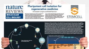 挂图Pluripotent Stem Cell Biology Nuclear transfer, reprogramming and cell fusion to induce pluripotency in somatic cells
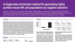 科学海报Cell Enrichment to Obtain Mouse NK Cell Populations By Negative Selection
 抗人TNF-α抗体,clone MT15B15 抗人TNF-α的小鼠Monoclonal IgG1抗体
 抗小鼠CD25抗体,clone 3C7 大鼠Monoclonal IgG2b抗体,抗小鼠CD25
 StemSpan™淋系祖细胞扩增添加物(10X) 用于人CD34+细胞扩增及分化为淋系祖细胞的添加物
 沪公网安备31010102008431号