
 EasySep™小鼠TIL(CD45)正选试剂盒


搜索结果: 'methocult media formulations for human hematopoietic cells serum containing'
-

产品类型:
产品号#:
19155
19155RF
产品名:
-
产品类型:
产品号#:
05850
05857
05870
05875
85850
85857
85870
85875
产品名:
mTeSR™1
mTeSR™1
-
产品类型:
产品号#:
18554
18554RF
18564
18564RF
产品名:
-
产品类型:
产品号#:
20164
100-0047
产品名:
RoboSep™ 缓冲液 2
EasySep™ Release 人PSC来源神经嵴细胞正选试剂盒
-
产品类型:
产品号#:
04230
84434
84444
产品名:
MethoCult™ H4230
-
产品类型:
产品号#:
21000
20119
20155
15862
15862RF
产品名:
RoboSep™- S
RoboSep™ 吸头组件抛光剂
RoboSep™分选管套装(9个塑料管)
-
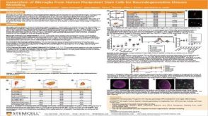 科学海报Generation of Microglia From Human Pluripotent Stem Cells for Neurodegenerative Disease Modeling
产品类型:
Conference:
ISSCR Toronto 2019
产品号#:
产品名:
发布日期: 12/12/2019 -
 实验方案How to Co-Culture Human Airway Epithelial and Immune Cells for RSV Infection
产品类型:
研究方向:
免疫学,呼吸系统研究,传染病
产品号#:
产品名:
-
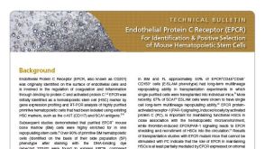 技术公告Endothelial Protein C Receptor (EPCR): A New Marker for Identification and Positive Selection of Mouse Hematopoietic Stem Cells
产品类型:
细胞类型:
造血干祖细胞
产品号#:
18556
18556RF
18554
18554RF
18564
18564RF
17665
17665RF
17666
17666RF
17696
17696RF
产品名:
EasySep™小鼠生物素正选试剂盒II
RoboSep™ 小鼠生物素正选试剂盒II
EasySep™小鼠PE正选试剂盒II
RoboSep™ 小鼠PE正选试剂盒II
EasySep™小鼠PE正选试剂盒II
RoboSep™ 小鼠PE正选试剂盒II
发布日期: 01/09/2017

 沪公网安备31010102008431号